Quantcast
Channel: Dr. Jackie Te RN, MD – Health Fixit
Viewing all 48 articles
Browse latest View live

Facet Arthropathy

$
0
0

What is Facet Arthropathy ?

Facet arthropathy has been considered a leading cause of lower back pain for over 50 years. It rarely involves the spinal nerves but can cause great distress and discomfort.

The facet joints are those that help with the flexibility in the back. Their scientific name is zygapophyseal joints.  The word arthropathy specifically means diseased joint. Therefore facet arthropathy is diseased facet joints found in the back and neck which can be noted with or without inflammation or swelling. It also does not include infection.  (1,3,7)

In this article, you will read more about the facet joint anatomy, symptoms, causes, diagnosis process and treatments.

Facet joint Anatomy

The facet joints are true synovial joints and are made up of the following:

  • Joint space
  • Hyaline cartilage surfaces
  • Synovial membrane
  • Fibrous capsule
  • Nerve fibers
  • Substance P (inflammatory mediator) (1)

The following link shows a quick video with a good explanation of the anatomy of the facet joint: http://www.spine-health.com/video/facet-joints-video

see a side view of the spinal bones and joints.photoFigure 1 : A side view of the spinal bones and joints.
Image Source : www.niams.nih.gov

The initial studies of facet arthropathy were done by injecting saline into the facet cavity. This caused inflammation and similar pain to that experienced with arthropathy. Therefore assisting doctors to better diagnosis the issue at hand(11)

see a cross section view of a normal vertebra.picture
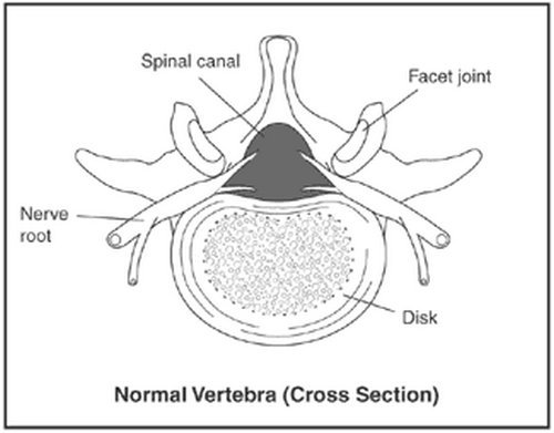
Image 2: The cross section view of a normal vertebra.
Photo Source : www.niams.nih.gov

Facet Arthropathy Symptoms

Lower back pain due to facet arthropathy is difficult to assess without using invasive techniques. But the following can be seen as common symptoms and may help with diagnosis:

  • Pain located in the lower back which radiates into the knee.
  • Usually, does not cause pain in the central back.
  • Pain could even radiate past the knee
  • Pain occurs a few times a month or as little as a few times per year
  • Tenderness to touch
  • Discomfort increases when bending backward more than when bending forward
  • Pain episodes that are recurrent
  • Muscle weakness
  • Numbness or tingling in extremities
  • In early stages may be noted as a stiffness in the mornings (1,3,4)

What is the difference between chronic and acute back pain?

  • Acute pain– Will begin to subside without treatment or with basic medications such as acetaminophen, aspirin, or ibuprofen.
  • Chronic pain– May or may not need surgery to go away. Usually does not get better with short term treatment and needs long-term physical therapy and behavioral changes.

How can a diagnosis be made?

Basically, your doctor will use the common symptoms stated above and may also use testing to rule out the following diagnosis:

  • Sacroiliac joint syndrome
  • Internal disk disruption syndrome
  • Lumbar spondylolisthesis and spondylosis
  • Chronic Pain syndrome
  • Lumbar compression fracture
  • Overuse injury
  • Fibromyalgia
  • Mechanical low back pain
  • Coccyx pain
  • Deep infection
  • Herniated disks
  • Fractures of the spine
  • Torn muscles
  • Acute intra-abdominal problems(1,3)

There are a couple studies that can be done to help determine facet arthropathy and disregard the above diagnosis. They consist of the following:

  • MRI Magnetic resonance imaging scans
  • Diagnostic blocks
  • CT scan
  • X-rays  (1,3)

lumbar spine.imagePicture 3 : A clear depiction of the lumbar spine which has faucet articulation issues.
Photo Source : naturalcuredoctor.com

What Causes Facet Arthropathy?

There could be many causes noted and each of these causes having their specific initiation point. The following is not a comprehensive list of possible causes for facet arthropathy:

  • Microtrauma
  • Osteoarthritis
  • Synovial capsule distention and inflammation
  • Rheumatoid arthritis
  • Wear and tear
  • Excessive lifting
  • Poor posture
  • Poor body mechanics when lifting or bending
  • Obesity
  • Sports that cause overuse (1,3,4)

Locations for Facet Arthropathy

Facet arthropathy can be found in the neck or in the back. If it is noted in the neck joints it is called cervical facet arthropathy. In the case that it is the lower back it would be called lumbar facet arthropathy.

Sometimes the specific vertebrae affected will be noted as part of the diagnosis and may appear as the following:

  • Facet arthropathy l5-s1
  • Facet arthropathy L2
  • Lumbar facet joint


The spinal areas are marked by their names.image

Image 4 : In this image, the spinal areas are marked by their names.
Picture Souece : www.simplebackpain.com

Types of Facet Arthropathy

Lumbar facet arthropathy

Lumbar facet arthropathy is the degeneration of the lumbar joints. This could be called L1-L5 as mentioned in the above section. This is due to poor body mechanics and can radiate to the areas noted in the above picture.

Cervical facet arthropathy

Cervical facet arthropathy is degeneration in the joints of the neck area. This could be called C1-C7. This could have many causes ranging from poor body mechanics to blunt trauma injury. This pain could radiate into the arms and chest.

Degenerative facet arthropathy

Degenerative facet arthropathy is just another way to describe the actions taking place with facet arthropathy. It may be used as a full in depth diagnosis.

Hypertrophic facet arthropathy

Hypertrophy is the increase of an organ or area of the body which is abnormal. In this situation it would be a section of the facet joint. This will cause pain in the area near the specific joint affected.

Bilateral facet arthropathy

Bilateral means both side, knowing this helps explain that bilateral facet arthropathy means that the degeneration is happening to both sides of the joint.

Thoracic facet arthropathy

thoracic facet arthropathy is degeneration in the joints of the thoracic (chest or upper back) area. This could be detailed as T1-T12 and can cause radiating pain to the arms, chest, and lower back. (1, 2, 6, 7)

Treatment

Surgical intervention is not needed for facet arthropathy. The following are other options of treatment. T(1,3)

Medications

Opioids

  • Tramadol
  • Codeine
  • Tricyclic antidepressants
  • Serotonin-norepinephrine reuptake inhibitors
  • Gabapentin
  • Pregabalin
  • In severe cases – morphine (5)

Facet joint injections

  • Steroid injections
  • Radiofrequency ablation
  • Intra-articular joint injection (1)
  • This link leads to a video which vividly explains the joint injections: http://www.spine-health.com/video/facet-joint-injections-procedure-video (3,5)

Facet joint Arthropathy exercises

Physical therapy

  • Traction
  • Behavioral modification (9)
  • Good posture is always helpful
  • Heat applications
  • Changes in routines
  • Sometimes anti-inflammatory medications can help with the pain. (3,5)

Prevention

Facet arthropathy is normally considered a mechanical problem due to misuse of the joints. The best prevention is as follows.

  • Good posture while sitting or standing
  • Proper body mechanics while lifting or bending
  • Maintaining flexibility through exercise
  • Flexion
  • Extension
  • Stretching (9)

Eating a healthy diet which includes

  • Good source of calcium
  • Good source of vitamin D

Exercise which increases strength in the back

  • aerobics (9)

Risk factors for Facet Arthropathy

There have been no studies to show that race or sex have any risk factors for facet arthropathy. Although some studies do show that age can cause significant risk.  This study showed 18% chance of people between the ages of 31-40 and 44% change in people between the ages of 51-60. (1)

Conclusion

In this article, we discussed the various treatments, causes, and prevention of facet arthropathy. The best way to prevent and help with pain due to facet arthropathy is proper posture and healthy lifestyle. Be sure to always seek medical attention if the pain is severe.

References:

  1. http://emedicine.medscape.com/article/310069-overview
  2. http://www.mayo.edu/pmts/mc5500-mc5599/mc5520-0116.pdf?_ga=1.18379641.1717306767.1484667538
  3. http://www.spine-health.com/conditions/arthritis/symptoms-and-diagnosis-facet-joint-problems
  4. https://www.laserspineinstitute.com/back_problems/facet_disease/articles/facet_joint_arthropathy/
  5. http://prc.canadianpaincoalition.ca/fr/facet_arthropathy.html
  6. http://www.fmcpaware.org/h-l/lumbar-facet-arthropathy
  7. https://www.reference.com/health/severe-facet-arthropathy-958b8055916b1907
  8. https://en.wikipedia.org/wiki/Arthropathy
  9. https://www.niams.nih.gov/Health_Info/Back_Pain/default.asp#4
  10. Essentials of Physical Medicine and Rehabilitation By Walter R. Frontera, Julie K. Silver, Thomas D. Rizzo Jr. https://books.google.com.mx/books?id=1sXsAwAAQBAJ&pg=PA233&dq=facet+arthropathy&hl=en&sa=X&redir_esc=y#v=onepage&q=facet%20arthropathy&f=false
  11. Atlas of Image-Guided Intervention in Regional Anesthesia and Pain Medicine By James P. Rathmell https://books.google.com.mx/books?id=DZhSxcok7poC&pg=PA82&dq=facet+arthropathy&hl=en&sa=X&redir_esc=y#v=onepage&q=facet%20arthropathy&f=false

Hypervolemia

$
0
0

Hypervolemia Facts

Hypervolemia, also known as fluid overload, is an excessive increase in the volume of fluid volume. It is an abnormal increase in blood plasma volume, which causes an elevated blood volume. [4, 2]

The condition involves expansion of the volume of extracellular fluid and covers the interstitial and intravascular space. The cause can be the overproduction of the isotonic fluid that is composed of sodium and water. [1,2, 3,6]

Hypervolemia and hyponatremia

Hyponatremia is a condition caused by abnormally low levels of sodium in the body. As an electrolyte, it regulates the amount of water in and around the cells. Hyponatremia causes the rise of water levels causing the cells to swell and retain water. [2, 4, 6, 7, 8]

Signs and Symptoms

One or more of these signs and symptoms may manifest when there is excess plasma volume:

  • Increased rate of respiration and shortness of breath. This is caused by the decreased number of red blood cells. Once blood is diluted, the body compensates it by an increase in respiration to enable all the cells to get enough oxygen.
  • An increase in the pulse rate, which is caused by the overload on the circulatory system and elevated contractility of the cardiac muscles.
  • Central venous pressure (CVP), pulmonary artery pressure (PAP) and shift of fluids from plasma cause edema in the ankles and the legs.
  • An increase in the volume of fluids among the lung pleural spaces causes labored and difficulty in breathing, a condition called pulmonary edema. The collection of the fluid in the lungs also causes paroxysmal nocturnal orthopnea (difficulty of breathing when lying down) and dyspnea (difficulty in breathing).
  • Crackling when auscultated due to the increase in hydrostatic pressure in the pulmonary arteries
  • Moist skin as the body tries to eliminate the excess fluid through sweating
  • Jugular vein distention caused by increased volume of the blood and the preload.
  • Excessive and rapid weight gain because of the building up of the extracellular fluids. There is also fluid buildups in the abdomen as the fluid drains from different parts of the body.
  • The feeling of a third heart sound, also known as S3 gallop during auscultation due to rapid filling of the articular part of the heart during systole and overloading of the ventricular part during diastole.
  • There is also an abnormality in the renal function that leads to specific alterations of gravity, change in the levels of the electrolytes, oliguria and azotemia.
  • Neurological changes such as change of the mental status characterized by lethargy or confusion, anxiety and restlessness. [2,3, 5,8,9,10, 11]

Hypervolemia and hypertension

Circulatory overload and an increase in the mean arterial pressure causes hypertension along with pulmonary artery pressure (PAP) and central venous pressure (CVP).  [1, 2, 3, 4, 5, 7, 11]

Hypervolemia and pregnancy

Pregnancy-induced hypertension called pre-eclampsia is common in women during their gestation period. It characterized by edema and presence of proteins in the urine. The expectant mother should get treatment for fluid retention promptly. [1, 3, 5, 10]

Hypervolemia pathophysiology

  • In the normal function, the body creates processes in which it enhances the electrolyte equilibrium, relinquishes, and compensates fluid. The process is achieved through the help of such hormones as aldosterone, antidiuretic hormone ADH and atrial natriuretic peptide ANP. The hormones influence the release of sodium and water that are essential to the body from the nephrons of the kidneys.
  • Here is an illustration of the fluid balancing mechanism. If an electrolyte is in higher concentration within the cells than the outside, fluids will move from outside the cells to inside the cell. If the electrolyte concentration outside the cells is higher than on the inside of the cell, the fluid moves outside. This body mechanism maintains the normal concentration of the electrolyte and balance of the fluid. [1, 3, 10, 11]
  • There may be cases of hypervolemia when they are cases of elevation of volume levels in the intramuscular space. The cause of the condition could be due to the shifting of the fluids from interstitium to the plasma, the excessive occurrence of intravenous fluids reduced water and sodium excretion and chronic stimuli on the renal part of the kidney causing the conservation of both the elements. [1, 3]
  • The elevation of the fluids in volume overloads the circulatory systems and leads to abnormally amplified contractility of the cardiac muscles, increases the mean artery pressure MAP and elevates the capillary hydrostatic pressure. The latter action leads to a shifting of the fluids to the interstitial space, which results in edema.  [1,2,3,4,5,10,11]
  • If the case of hypervolemia gets severe or in case the patient suffers from a history or cardiac dysfunction, the body’s compensatory mechanisms may not work. Moreover, he or she may experience a shortfall of urinary excretion of water and sodium. In a worse case, the aldosterone and antidiuretic hormone may not be stopped from causing an elevation of mean arterial pressure. The result is usually pulmonary edema, which is followed by heart failure. [1, 3,]

 Causes of Hypervolemia

  • Excessive intake of fluid or sodium
  • Intravenous (IV)therapy
  • Hypervolemia may occur if an intravenous fluid is administered in a larger volume or rapid rate than directed by the doctor.
  • When a wrong intravenous fluid is administered, it may alter the sodium levels in the blood.
  • Use of hypotonic fluids on perioperative patients who are undergoing an intravenous therapy [1,3]
  • Body’s reaction to transfusion when a large volume of blood products are infused or the process if infusion done rapidly [3]
  • Excessively taking of dietary sodium  [1,2,3,4, 5, 11]

Retention of water and sodium

Heart problems

  • Acute and chronic heart diseases
  • Congestive heart failure; where the heart is unable to pump the blood effectively throughout the body. The blood backs up in veins causing an increase in fluid volume. [1,3,5]

Kidney disorders

  • Glomerulonephritis that makes the kidneys unable to excrete excess fluids from the body.
  • Nephritic syndrome that causes the deficiency in as protein called albumin. The deficiency leads to transfer of fluids from the blood to the body tissues, which ends in edema. The kidney on the other hand, tends to retain more fluid and salt. [7,8]
  • Liver cirrhosis also contributes to the overload of the fluid as it leads to increase in plasma levels [1,2, 3]
  • Certain medications such as chemotherapy drugs and corticosteroid therapy drugs [1,2,3]
  •  Low protein intake [3]
  • The body’s reaction to stress such as after an injury of surgery. Head injury patients all experience increased antidiuretic hormone levels that lead to sodium and water retention. [3,8]

Shift of fluids into intravascular space

  • In burn patients during fluid remobilization
  • Intravenous infusion of plasma proteins [ 1,3]


Leg edema because of a result of capillary leak syndrome
Source: Wikimedia

Diagnostic tests and lab values

Below some of the common laboratory tests used for accessing a person’s hydration status

  • Hematocrit test – This test measures the percentage of the blood that is made up of red blood cell. The test is often a part of complete blood count. Hematocrit decreases at the increase of hydration
  • Normal adult male values; 40.7 % to 50.3 %
  • Normal female values : 36.1 % to 44.3 %
  • Sodium test – The test measures the amount of sodium in the blood and is often done when the doctor suspects electrolyte imbalance. Overhydration causes hyponatremia, which are low amounts of sodium in the blood.  Normal values in adults is 135-145 mEq/L
  • BUN (Blood Urea Nitrogen Tests) – The test measures the amount of nitrogen and urea in the blood as part of kidney function evaluation. BUN decreases with overhydration. Normal adult values: 10-20mg/dl
  • Serum Osmolality Test – The test seeks to determine the number of solute in the blood or serum sample. It is a common test in the evaluation of hyponatremia (loss of sodium). Osmolality decreases with overhydration. [6, 8]

Pitting edema LEG
Pitting edema before and after the application of pressure on the skin. The action shows the extent of swelling in the leg.
Source: Wikimedia

Treatment of Hypervolemia

There are several treatments and nursing interventions that can manage the problem as listed below

  • Medication – Patients are given diuretics to help in the excretion of the excess fluids while heart failure patients are given inotropes
  • Modification of diet – The doctor may recommend a low salt diet for hypervolemia patients to ease the workload on the kidneys.
  • Isolated ultrafiltration – The process uses an extracorporeal filter for the elimination of isotonic fluid. It is effective in the removal of excess sodium from the body. [1, 2,3,4,5, 9, 10, 11]

References

  1. http://www.healthresource4u.com/hypervolemia.html
  2. http://www.ihealthblogger.com/2013/07/Hypervolemia-Definition-Symptoms-Diagnosis-and-Treatment.html
  3. http://howshealth.com/hypervolemia/
  4. Mayoclinic.org
  5. http://patient.info/doctor/fluid-overload
  6. Physio-pedia.com. Fluid Excess/Intoxication – Physiopedia, universal access to physiotherapy knowledge. [Internet]. 2015 [cited 22 December 2015].
  7. Weir M. Hypervolemia and Blood Pressure: Powerful Indicators of Increased Mortality Among Hemodialysis Patients.
  8. Androne AS e. Relation of unrecognized hypervolemia in chronic heart failure to clinical status, hemodynamics, and patient outcomes.  – PubMed – NCBI [Internet]. Ncbi.nlm.nih.gov. 2015 [cited 22 December 2015].
  9. Merck Manuals Professional Edition. Hyponatremia – Endocrine and Metabolic Disorders [Internet]. 2015 [cited 22 December 2015].
  10. Hypervolemia.com
  11. Drjohnivanoffmdtulsa.com. Fluids Hypervolemia And The Heart

How to increase platelet count

$
0
0

Platelets circulates within the body and when they recognize damaged blood vessels, they bind together to clot the blood and prevent further bleeding. Platelet is the smallest blood cell and is only visible under the microscope.

It is called platelet because it looks like small plates. During injury or damage, the blood vessels will send signal to the platelets and they will then travel to the area of damage and transforms into active form. Once the platelets are in their active form, they will grow long tentacles, which resembles like an octopus or spider.


A group of food that can significantly increase the number of platelets in the body.picture

Image 1 : A group of food that can significantly increase the number of platelets in the body
Photo Source : www.medindia.net

A patient with a petechial rash.imagePicture 2 : A patient with a petechial rash
Photo Source : mayaclinic.in

Healthy platelet count – Normal range

A normal platelet count ranges between 150,000 and 450,000 per microliter of blood. If there is more than enough level of platelet, the patient is suffering from thrombocytosis. If the platelet is abnormally low, the patient is suffering from thrombocytopenia. The number of platelet can be easily checked through complete blood count test (CBC).

Symptoms of low platelet in the blood

  • Easy bruising
  • Rash, specifically a number of petechiae or also known as purpura
  • Frequent bleeding from the nose and gums
  • Bleeding of the gastrointestinal tract as seen on melena (blood in the stool)
  • Blood in the urine
  • Bloody or dark vomit

What causes a low platelet count? The platelet drops if there are circumstances that prevent the body from producing platelets. It could be due to inherited condition, side effects of medications, kidney infection, too much level of alcohol in the blood, adverse effect of chemotherapy, and viral diseases like Dengue fever.

However, there are a few circumstances when the number of platelets goes a bit lower such as during pregnancy and when women over their menstrual cycle.

An abnormally low or severely low platelet should be addressed the soonest time possible because it can put the patient at risk for bleeding. The bleeding is even more possible if the platelet count goes below 50,000 per microliter. You could bleed even if you have no injury at all. If the platelet count is below 5,000 per microliter, you could suffer from spontaneous bleeding. It can put your life in danger.

Diagnosis and Treatment

Before implementing measures on how to increase platelet counts, you should first come up with the right diagnosis. To correctly diagnose the condition of the patient, the doctor will ask questions and will conduct a thorough physical examination.

A complete blood count is ordered to check the platelet concentration. In a severe condition, a bone marrow biopsy is needed.

How to increase platelet count?

There are various ways to increase platelet count. If you want to know how to increase platelet count fast, then you need to find out the cause of low platelet and severity of condition. If the platelet is mildly low, the doctor will monitor your condition and will refrain you from doing things that could worsen your condition.

You should avoid contact sports and activities that could lead to bruising or bleeding. Furthermore, you have to limit if not totally avoid alcohol consumption. Any medications that can affect the level of platelet should be stopped. Examples are aspirin and ibuprofen.

If the level of platelet is severely low and you are looking for ways on how to increase platelets instantly, the solutions are as follows:

  • Blood/platelet transfusions
  • Stop the drugs that caused low platelet
  • Block the platelet antibodies by taking corticosteroids

How to increase low platelet count naturally?

Papaya

If you are looking for ways on how to increase platelet count during dengue or whatever the reasons are, then all you need is papaya. The fruits and leaves of papaya can increase the number of platelet in just a few days. Patients suffering from dengue fever will see an increase in their level of platelet after taking papaya leaves juice.

If you don’t like the taste of the leaves, then you can just eat a ripe papaya. You can also drink a glass of papaya juice. Add lemon juice for added vitamin C. drink papaya juice two to three times a day and your platelet will increase in no time.

Wheatgrass

It is one of the commonly used home remedies for low platelet. It will not only increase platelet count, but as well other blood components like haemoglobin, white blood cells, and red blood cells. Wheatgrass has a high level of chlorophyll, which has a molecular structure similar to that of the haemoglobin molecule. A cup of wheatgrass juice a day can significantly increase the level of platelet.

Pumpkin

How to increase platelet count through diet? The answer is pumpkin. You need to add a lot of pumpkin to your diet. It can improve the level of platelet. Pumpkin is rich in vitamin A, a nutrient useful for the proper development of platelet.

Pumpkin does a very good job in regulating protein in the cells, which plays a vital role in increasing the level of platelet. You can drink a glass of pumpkin juice two to three times a day. You can also include pumpkin in your favorite dishes and baked goodies.

Spinach

Spinach is rich in vitamin K, which is useful in the production of platelet. Vitamin K plays a very important role in the blood clotting process. If your platelet is low, you should eat plenty of spinach to avoid bleeding. Enjoy spinach by adding in vegetable salads. You can also make spinach soup, smoothie, or as a side dish. You can also drink spinach juice two to three times a day.

Vitamin C

Increasing your intake of foods rich in vitamin C can help improve the level of platelet. Vitamin C is a potent antioxidant. It plays a vital role in preventing free-radical mediated damage of the platelets. Examples of foods high in vitamin C content includes oranges, lemons, kiwi, cantaloupes, spinach, broccoli, and bell peppers. If you don’t like the taste of these foods, then your best option is vitamin C supplement.

Indian Gooseberries

Indian gooseberries can help elevate the level of platelet. It boost the immune system and improve the level of platelet. The ideal way of taking Indian gooseberries is early in the morning in an empty stomach. Eat at least three to four gooseberries.

Pomegranate

If your platelet is low, then you should eat a lot of pomegranate. It is rich in iron, which is helpful in improving the level of platelet in the blood. Pomegranate can be eaten raw. You can also squeeze and drink the juice.

Eat a lot of pomegranate for two weeks and you will see a significant improvement in your platelet and energy level.

Milk

The calcium in milk helps the body in regenerating a significant amount of platelets. Calcium works hand in hand with vitamin K and fibrinogen (milk protein) in improving the level of platelet in the body. It strengthens the ability of the blood to form clots. Other foods rich in calcium are yogurt, cheese, and organic dairy products.

Lean Protein

It is rich in zinc and vitamin B12. What lean protein does is it reverses the effect of low platelet. Examples of foods rich in lean protein are turkey, fish, oysters, and chicken.

References:

  1. www.thrombocyte.com
  2. www.top10homeremedies.com
  3. www.wikihow.com
  4. www.rapidhomeremedies.com
  5. www.quora.com
  6. www.livestrong.com
  7. https://en.wikipedia.org
  8. www.hopkinsmedicine.org
  9. www.healthline.com
  10. www.medicalnewstoday.com

Dactylitis

$
0
0

Dactylitis pertains to inflammation of the joints in the fingers and toes.

What is sausage finger syndrome? It is sometimes called a sausage toe or finger because the digits look like sausages because of inflammation. In severe cases, it can make your fingers rigid that you can’t even make a fist.

If you are suffering from dactylitis, it can indicate other serious health issues. You could be suffering from arthritis, sickle cell anemia, or ankylosing spondylitis. (1)

Dactylitis causes

There are various reasons for dactylitis and the common causes include the following:

  • Dactylitis Psoriatic Arthritis – It is an autoimmune disease causing the immune system to attack healthy tissues. It causes pain and inflammation, especially to the joints and tendon sheaths.
  • Dactylitis Rheumatoid Arthritis – Rheumatoid arthritis is an autoimmune disorder, which causes the immune system to attack the joints. As a result, the patient experiences severe pain around the joints. The joints get inflamed and due to severe swelling, the fingers and toes look like sausage. It affects the hands, wrist, feet, knees, elbows, and ankles. (1, 2)
  • Reactive Arthritis – This is caused by reaction in some parts of the body. It could be an infection in the urinary tract, genitals, or intestines. Reactive arthritis is usually caused by some types of bacteria such as salmonella, Yersinia, shigella, and campylobacter. Reactive arthritis causes stiffness, urinary tract problems, and inflammation of the eyes.
  • Dactylitis Sickle Cell – Dactylitis is the first symptom of sickle cell disease, especially in babies. On the positive note, Dactylitis does not cause any damage to the child’s body. (3)

Dactylitis in infant with sickle cell disease.phototImage 1 : Dactylitis in infant with sickle cell disease
Picture Source : img.medscape.com

Sausage-like fingers in a patient with psoriatic arthritis.image

Picture 1 : Sausage-like fingers in a patient with psoriatic arthritis
Photo Source : www.healthline.com

What are the symptoms of dactylitis?

The symptoms can appear abruptly or may take a few days before they start to develop. The symptoms vary depending on the cause. The primary symptoms include swelling and pain in the digits, the affected part of the body is difficult to move. Sometimes, you cannot move your digit or even make a fist. The joints feel warm.

Accompanying symptoms include fever, shortness of breath, fatigue and visual disturbances. Dactylitis caused by psoriatic arthritis causes the individual digits to swell differently. On the other hand, rheumatoid arthritis causes symmetric pattern of swelling in various parts of the body. (3, 4)

When you should call out for help?

If you are having an extremely high fever and unbearable pain, then you should immediately ask for help. You should see your doctor right away if the pain won’t go away despite taking pain reliever at home. If the problem is not treated immediately, it could lead to severe conditions like pulmonary hypertension, chronic renal failure, decrease immune reaction, secondary infections, and stroke. (5)

How is dactylitis diagnosed?

A lot of factors could lead to dactylitis. To accurately diagnose the condition of the patient, the doctor will conduct a series of test. Physical assessment and interview of past medical and surgical history will be done to rule out what could possibly be causing dactylitis.

The doctor will thoroughly assess the location of the pain and swelling, particularly on the joints, toes, and fingers. An imaging test such as x-ray and magnetic resonance imaging (MRI) will be ordered to accurately view the affected parts of the body.

If the doctor is suspecting gout or rheumatoid arthritis, he will order laboratory tests like joint fluid test and additional blood test. It is important to correctly and immediately diagnose the condition that causes dactylitis. Failure to do so could lead to permanent joint deformity and eventually loss of function of a particular body parts. (5, 6)

Dactylitis Differential Diagnosis

Dactylitis or sausage-shaped fingers could indicate a particular type of psoriatic arthritis minus the rheumatoid factor activity. If the joint pain progresses along the phalanges of fingers along with soft tissue swelling, it is a confirmatory assessment that the patient is indeed suffering from dactylitis. (7)

Dactylitis Treatment

The treatment approach for dactylitis varies depending on the underlying cause. If it is caused by psoriatic arthritis, unfortunately there is no available cure. The best thing to do is to manage the symptoms or so-called symptomatic relief.

The doctor will prescribe non-steroidal anti-inflammatory drugs as well as disease-modifying anti-rheumatic drugs. These drugs can alleviate pain and somehow decrease the inflammation. They also prevent damage to the joints. (7)

If the cause of dactylitis is rheumatoid arthritis, the doctor will prescribe a variety of medications such as corticosteroids, disease modifying anti-rheumatic drugs, JAK (Janus Kinase) inhibitors, and genetically-engineered proteins. If the condition is extremely severe, a joint replacement surgery will be performed. However, this should be the last resort. It would only be performed if all other remedies failed.

If the dactylitis is caused by reactive arthritis, the doctor will treat it using antibiotics, corticosteroids, non-steroidal anti-inflammatory drugs, and rheumatoid arthritis drugs. The patient is also advised to have physical therapy and exercise to significantly improve the functions of the joints and reduce stiffness. (8)

Other forms of dactylitis

  • Blistering distal dactylitis – It is an inflammation on the distal part of the fingers. It usually affects the children, especially between six months and four years. It affects the fat pad of the finger. There is pain and inflammation, which makes it extremely difficult to move your fingers. It is caused by group A haemolytic streptococcus and Staph aureus. The treatment approach for this type of dactylitis include warm compress, oral antibiotics, and incision and drainage.
  • Tuberculous dactylitis – It does not affect the flesh of the fingers and toes, but rather affect the bone structure of the hands and feet. It causes severe pain, especially in the short tubular bones. (8,9)

Pain management for dactylitis at home

Mild episodes of dactylitis can be treated at home. In case of pain and discomfort, the patient should take over the counter pain reliever such as ibuprofen. You should also increase your intake of fluids, especially water and fresh fruit juices. A warm compress on the swollen area constricts the blood vessels, which helps alleviate pain and discomfort. (10)

References:

  1. http://www.healthline.com
  2. https://en.wikipedia.org
  3. www.ncbi.nlm.nih.gov
  4. diseasespictures.com
  5. www.webmd.com
  6. https://patient.info
  7. www.mdedge.com
  8. howshealth.com
  9. www.epainassist.com
  10. www.rightdiagnosis.com

TNF Alpha Inhibitors

$
0
0

TNF Alpha Inhibitors or Tumor Necrosis Factor inhibitors are a classification of drugs used to treat inflammatory conditions like inflammatory bowel disease, psoriatic arthritis, rheumatoid arthritis, juvenile arthritis, psoriasis, and ankylosing spondylitis.

What these drugs do is they stop the progression of the disease and reduce the inflammatory response by targeting the tumor necrosis factor. (1)

What is TNF?

Tumor Necrosis Factor (TNF) is a type of protein created by white blood cells in response to infection and inflammation. TNF is useful because it heightens the immune response of the body. However, TNF can activate various bodily reactions including cancer cell growth. In a healthy person, if the number of tumor necrosis factor goes out of control, it is blocked naturally by the body.

However, people with rheumatic disease who suffer from excess TNF could suffer from severe inflammation and other associated symptoms. It affects not just the inflammation in the joints, but as well in the gastrointestinal tract and skin. (1, 2)

There is a vast TNF alpha inhibitors list, but the US Food and Drug Administration approved only six TNF inhibitors for the treatment of rheumatic conditions. TNF alpha inhibitor drugs are quite costly. To somehow reduce the cost, patients with mild to moderate disease are treated with methotrexate before switching to TNF inhibitor. (2)


Image 1 : Tumor necrosis factor alpha inhibitor biologics and mode of action
source: medscape.com

Turmeric, one of the spices packed with health benefits.imagePicture 2 : Turmeric, one of the spices packed with health benefits.
Photo Source : cdn.authoritynutrition.com

A plant loaded with health benefits including the ability to block TNF alpha..photo

Image 3 : A plant loaded with health benefits including the ability to block TNF alpha.
Picture Source : s3.amazonaws.com

TNF alpha inhibitors mechanism of action

TNF alpha inhibitor is useful in the treatment of various inflammatory conditions. TNF alpha inhibitors for rheumatoid arthritis are tested and proven effective. In fact, they are more effective than traditional anti-inflammatory drugs. However, clinical trials and post marketing surveillance showed that TNF alpha inhibitors have adverse effects. (2, 3)

Tumor necrosis factor alpha plays an important role in the inflammatory and pain pathways. By inhibiting the action of TNF, the inflammatory response of the body will be reduced. This mechanism of action of TNF alpha inhibitor makes it perfect for the treatment and management of autoimmune conditions and inflammatory diseases.

TNF alpha inhibitor drugs can significantly decrease the pain, improves the function of the body, and improve the joint damage in people with rheumatoid arthritis.

Before starting TNF alpha inhibitors, a person should firsts screen for HBsAg. People with HBsAg should receive prophylaxis. TNF causes immunosuppressive response, which could lead to reactivation of infections like hepatitis B and tuberculosis. (4, 5)

Side effects of TNF alpha inhibitors

Tumor necrosis factor given to the patient through injection can cause skin reaction. There is a localized rash on the injection site and the patient complains of itching or burning sensation. This usually last for a week. TNF alpha inhibitors can also put the patient at risk for various types of infections, especially fungal infections, tuberculosis, and hepatitis B infection.

Hence, it is highly recommended for the patient to be tested for TB before starting TNF alpha inhibitor therapy. The testing is done via blood test and skin test. TNF alpha inhibitors when used for a long period of time can put a patient at risk for cancer, especially skin cancer and lymphoma.

Those with multiple sclerosis or those with history of MS shouldn’t take TNF alpha inhibitors. People with cardiac disease should completely avoid TNF alpha inhibitor because it can further complicate the disease. (6)

What to keep in mind?

If the patient is currently on TNF alpha inhibitor and he suddenly has high fever, you should stop the drug right away. Patients who are on antibiotics for infection should not receive TNF alpha inhibitor too. The infection should completely go away before restarting TNF alpha inhibitor.

If a patient is planning to get vaccination while on TNF alpha inhibitor, he should first inform his doctor. Live vaccines should be completely avoided, although some vaccinations are safe. (7)

TNF alpha inhibitors could eat up a huge portion of your budget. The cost could be more than $10,000 a year. On the positive note, the expenses for TNF alpha inhibitor is usually covered by most health care insurance plan.

Natural TNF alpha inhibitors

As mentioned above, TNF alpha inhibitors are expensive. If you are looking for ways to improve your health without having the need to spend more than what you can, then you should switch to natural TNF alpha inhibitors. These include the following:

  • Turmeric – One of the common TNF alpha inhibitors is turmeric. It is an herb commonly used in Indian cooking. It is readily available in the leading supermarket. Turmeric contains curcumin, a chemical used to alleviate inflammation. It inhibits tumor necrosis factor. If you are the type of person who does not like the taste of turmeric, then don’t worry as there is now curcumin in capsule form. You can easily purchase it at the leading drug stores. (7, 8)
  • Milk Thistle/Holy Thistle – It is a type of plant primarily used to protect and treat liver-related diseases. The primary component of milk thistle is silymarin, which is high in antioxidant and anti-inflammatory. Silymarin can block tumor necrosis factor from activating nuclear transcription factor, which controls the inflammatory response and development of cancer cells. To benefit from the TNF alpha inhibitor effect of milk thistle, a person should consume about 280 mg to 450 mg of milk thistle a day.
  • Stinging Nettle Leaf Extract – This is one of the best herbal remedies for chronic inflammatory diseases. The leaves of stinging nettle can be brewed into a tea. The extracts can be taken as a replacement for water. (6, 9)
  • Green Tea – Natural green tea contains catechins, a potent chemical that plays a vital role in alleviating inflammatory response of the body. Drink 300 mg to 600 mg of green tea a day to enjoy its anti-inflammatory benefits. (10)

Tumor necrosis factor alpha inhibitors have been a huge help in the treatment and management of inflammatory diseases, especially arthritis. Millions of people all across the globe are benefiting from TNF alpha inhibitor therapy.

References:

  1. http://www.arthritisresearchuk.org
  2. www.naturalmedicinejournal.com
  3. https://en.wikipedia.org
  4. https://www.rheumatology.org
  5. www.healthline.com
  6. www.medscape.com
  7. www.life-enhancement.com
  8. www.greenmedinfo.com
  9. www.gov.uk
  10. www.medbullets.com

Blood in the Stool

$
0
0

Noticing an occult blood in stool can be very frightening. It can signal a serious problem and so it is important to have it checked right away. There are a lot of reasons for blood in stool. The amount of blood varies from one person to another.

Some people experience a bit of blood while others have a large amount of blood in the stool. A fresh blood in stool medical term is hematochezia. On the other hand, a dark stool or medically known as melena can suggest internal bleeding. (1, 2)

What are the associated symptoms?

There is mucus and blood in stool. Other associated symptoms include:

  • Vomiting
  • Abdominal pain
  • Diarrhea
  • Weakness
  • Weight loss
  • Difficulty of breathing
  • Palpitations (2)

Picture of blood in stool

An image of the gastrointestinal tract outlining the possible location of blood.picture

Image 1 : An image of the gastrointestinal tract outlining the possible location of blood
Photo Source : www.epainassist.com


An anatomical representation of the gastrointestinal tract.image

Picture 1 : An anatomical representation of the gastrointestinal tract
Photo Source : s-media-cache-ak0.pinimg.com

colonoscopy.photo

Image 3 : A picture that shows how colonoscopy is done
Picture Source : d2if36jingo4w0.cloudfront.net

What can cause blood in stool?

What does blood in your poop mean? Blood in the stool could suggest that something is wrong in your digestive tract. There could be a possibility of bleeding in the GI tract. Most of the time, the amount of blood is small that it can easily be detected on fecal occult test. If the bleeding is in large amount, then it could suggest a serious problem and warrants immediate medical attention. Possible reasons for blood in stool include the following:

  1. Piles/Hemorrhoids – the blood vessels in the rectal area are swollen. If your poo is hard, there is a possibility of bleeding. (2)
  2. Diverticular disease – Can you bleed with diverticulitis? Diverticula are tiny pouches projecting the colon wall. A bleeding is possible if the diverticula gets infected. (3)
  3. Anal fissure – It pertains to small cut or tear in the lining of the anus. It can be due to passage of a hard and large tool. It can be very painful and causes bleeding.
  4. Colitis – It is the inflammation of the colon. It can be caused by inflammatory bowel disease. If proper medication and management are not given, the patient could suffer from gastrointestinal bleeding, which leads to the presence of blood in the stool.
  5. Angiodysplasia – It is a condition characterized by sensitive and abnormal blood vessels. Even the simplest pressure could lead to bleeding.
  6. Peptic ulcer – It is an open sore, which can be present in various parts of the GI tract such as the duodenum, stomach, and upper end of the small intestine. Peptic ulcer is usually caused by H. pylori, but it could also be an adverse effect of medications such as ibuprofen, aspirin, and naproxen. (4)
  7. Polyps or cancer – A polyp is a benign growth that once gets out of control, could grow bigger and bleed. Over time, it can lead to cancer. Colorectal cancer causes bleeding, which is responsible for the presence of blood in the stool. Colorectal cancer is one of the most common cancers in America. (4, 5)

Diagnosis and Treatment

To come up with the best blood in stool treatment, it is important to properly diagnose the condition. The doctor will thoroughly evaluate the bleeding in the stool. The color of the blood has a lot to do with the root cause of the problem.

A black, tarry stool is an indicator that there is a problem in the upper part of the gastrointestinal tract. If the color of the blood is bright red or maroon, it is an indicator that something is wrong in the lower gastrointestinal tract. It could be due to diverticulitis or haemorrhoids. (5)

Various diagnostic procedures are used to detect the root cause of the problem. They include the following:

  • Nasogastric lavage – This procedure tells exactly where the source of bleeding is. In this procedure, a tube is inserted into the stomach through the nose to remove the content of the stomach. If there is no blood in the upper GI tract, the source of bleeding could be in the lower GI tract. It could also be possible that the bleeding already subsided.
  • EGD (Esophagogastroduodenoscopy) – An endoscope is inserted through the mouth going down the esophagus, stomach, and duodenum. This procedure is done to accurately detect the source of bleeding.
  • Colonoscopy – It is somewhat similar to that of the EGD. The difference is that in colonoscopy, the scope is inserted through the rectum to have a clear view of the colon. It can also be used to collect a tissue sample for biopsy.
  • Enteroscopy – This procedure is specifically for the small intestines. The patient should swallow a capsule with a tiny camera, which transmits images to video monitor as it passes through the gastrointestinal tract.
  • Barium X-ray – It uses a barium, which is a contrast medium that makes the gastrointestinal tract visible on x-ray. The patient has the option to swallow the barium or have it inserted into the rectum.
  • Radionuclide scanning – A small amount of radioactive material is injected into the vein. To see the images of blood flow in the GI tract, a special camera is used. This can help detect the source of bleeding.
  • Angiography – A special dye is injected into the vein and its purpose is to make blood vessel visible on CT scan or X-ray.
  • Laparotomy – This is an invasive medical procedure in which the doctor opens and examines the abdomen. This test should be the last resort. It can only be done if all other diagnostic procedures fail to detect the root cause of the bleeding. (5, 6, 7,8)

Note: A laboratory test in the form of blood stool is ordered to watch out for any signs of bacterial infection, anemia, and blood clotting problems.

Treatment and Management

The primary goal of treatment is stop immediately stop the bleeding. One of the common approach is to inject a substance to the bleeding site and treat it with an electric current/laser. Some doctors prefer to clip the bleeding vessels. The doctor can also inject a special type of medicine into the blood vessels to immediately stop or at least somehow control the bleeding. (9, 10)

The remedy does not end in stopping the bleeding. The root cause should be determined so as to avoid the problem from happening again. As with the treatment, it varies depending on the cause of the problem. If it is caused by bacteria, an antibiotic is prescribed. If it is caused by colitis, an anti-inflammatory medication should be given to the patient. A cream or suppository is required if the bleeding is caused by haemorrhoids. On the other hand, if the bleeding is caused by polyps, a surgery is warranted.

There are simple measures that you can do to stop, control, and prevent bleeding. A blood in the stool can be prevented from happening again if you eat foods rich in fiber. This is helpful, especially if you are constipated. Constipation can aggravate haemorrhoids, which increases the chance of bleeding. If you have blood in the stool secondary to fissures, you can relieve the pain and bleeding by sitting in a warm bath. (9, 10)

References:

  1. www.webmd.com
  2. www.medicinenet.com
  3. www.mayoclinic.org
  4. www.womenshealthmag.com
  5. www.livestrong.com
  6. www.thrombocyte.com
  7. www.healthline.com
  8. https://en.wikipedia.org
  9. www.doctorshealthpress.com
  10. Everyone’s Guide to Cancer Therapy: How Cancer Is Diagnosed, Treated, and Managed Day to Day By Andrew Ko

Brachioradialis Pain

$
0
0

What is a brachioradialis muscle?

What does the brachioradialis do? Brachioradialis is one of the muscles in the forearms. It is also called Venke’s muscle. It extends from the lower part of the humerus down to the radius.

The humerus is the long bone in the upper arm and the radius is the long bone on thumb side of the forearm. Brachioradialis is both superficial and fusiform muscles.

Superficial muscles are located underneath skin and fat tissues while the fusiform muscles are skinny at their ends and thicker in the middle. They look like a spindle. (1, 2)

The brachioradialis muscle situated in the forearm.photo

Image 1: The brachioradialis muscle situated in the forearm.
Picture Source: seannal.com

Outlining the different muscles of the arms.image

Photo 2: Outlining the different muscles of the arms.
Image Source: seannal.com

The trigger point of brachioradialis muscle.photo

Image 3: The trigger point of brachioradialis muscle.
Picture Source: www.triggerpointtherapist.com

Functions of brachioradialis muscle

  • Flexion of the forearm (raising the forearm by bending the elbow).
  • Pronation of the forearm (rotating the forearm).
  • Supination of the forearm (rotating the forearm so the palm faces up). (1, 2)

Brachioradialis Nerves

The muscles in the body are connected to a nerve, which supplies electrical impulses to the muscles from the brain. The radial nerve is the one that sends nerve impulses to the brachioradialis muscles. It is a large nerve consisting of nerve fiber from C5 to C7. These are the nerves originating from the neck region of the spinal cord.

However, if the muscles in the forearm become extremely tight, a shooting pain can be felt in the forearm and elbow. This is referred as brachioradialis pain. Brachioradialis pain after lifting heavy objects is common. There are instances when the pain extends to the back of the hand and into the thumb and index fingers. Brachioradialis pain and swelling are usually felt when shaking hands, turning a door knob, using a screwdriver, and drinking a cup of coffee. (3, 4, 5, 6)

What cause brachioradialis pain?

  • Overexertion – It is one of the primary causes of brachioradialis pain. It is caused by overloading of the muscles for a long period of time. Eventually, the muscles become tender and painful. Manual labor could lead to overexertion of brachioradialis muscles and other surrounding muscles. Simple activities can also trigger brachioradialis pain such as typing on a computer for a long period of time and playing tennis. Frequent lifting, twisting, and holding things increase the stress in brachioradialis muscles.
  • Trauma/injury – An injury or trauma to the arm can lead to brachioradialis pain. A sudden fall from a high place or a blunt force increase the possibility of brachioradialis pain. The pain is sharp and intense. (5, 6)

How to distinguish brachioradialis pain?

  • A brachioradialis pain is felt along with muscle weakness.
  • The onset of pain is sudden.
  • The muscles of the arm get weak.
  • The pain is described as sharp, radiating, and piercing.
  • The origin of the pain is in the shoulder and upper arm.
  • The pain is felt on one side of the body.
  • The pain is felt on the front part of the elbow.
  • The patient is unable to completely straighten or bend the elbow because the pain gets severe when attempting to do so.
  • Simple activities intensify the pain.
  • When the tendon or elbow is touched or moved, you will hear a crackling sound.
  • The pain is temporarily relieved by pain killers but will recur again after a few hours.
  • The shoulder muscles get weak. In severe cases, the shoulder muscles become paralyzed.
  • The patient may complain of shortness of breath if the nerve near the diaphragm is affected. (6, 7, 8)

How to diagnose brachioradialis pain?

If you are experiencing brachioradialis pain, you should immediately consult your doctor. The doctor will conduct various diagnostic procedures and physical assessment to thoroughly check the condition of the patient.

The doctor will perform electromyography or nerve conduction studies to confirm the diagnosis of brachioradialis pain. It helps detect the extent of nerve damage. The strength and movement of the shoulders will be assessed too.

There are instances wherein the affected shoulders protrudes. Depending on the overall condition of the patient, the doctor will order for additional tests such as CT scan, MRI, and x-ray of the shoulders and neck. (8, 9, 10)

Brachioradialis Pain Treatment

A brachioradialis pain should be treated the soonest time possible. The focus of the treatment is to alleviate the pain, control the nerve damage, restoration of the functions of brachioradialis muscles, and improve the overall health. A brachioradialis tear can be improved using the right treatment modalities.

Rest

A painful brachioradialis muscle should be kept rested. After an injury, the muscle should be rested for at least 72 hours. You should apply ice with a 20-minute interval every two hours. This is to minimize swelling and inflammation. You should keep the affected area elevated, especially if it is caused by blunt injury. (3)

Brachioradialis Exercises

A brachioradialis tear can be improved using range of motion exercises. What are some exercises for brachioradialis? They include the following:

Isometric exercises

This involves contraction of the brachioradialis muscle for a specific timeframe. The patient should hold dumbbells while standing. Then, lift the weights at about three inches forward. Repeat the process until such time you will be able to see a significant improvement in your muscles. (1, 4)

Strength training exercises

Lifting heavy weights can help improve the condition of the brachioradialis muscle. However, you need to seek the advice of a physiotherapist to determine if you can now start lifting heavy weights. Exercises that target the brachioradialis muscles include hammer curl and reverse curl.

  1. Barbell curl – This is effective in training the brachioradialis muscles. Make sure that the weight of the barbell correlates with the strength of the arm muscles. The barbell should not be too light or too heavy.
  2. Dumbbell hammer curl – The barbell should be curled toward the shoulders. Performing dumbbell hammer curl exercise regularly will help regain the tone and tightness of the brachioradialis muscles minus the pain.
  3. Arm extension exercise – Keep the affected arm at the side and slowly straighten the elbow. Push down the forearm until such time you will feel a gentle stretch on the inside part of the elbow. (5, 8, 9)

References:

  1. https://www.belmarrahealth.com/brachioradialis-pain-symptoms-treatment-pain-relief-tips/
  2. https://www.muscle-joint-pain.com/trigger-points/trigger-point-self-treatment/brachioradialis/
  3. http://www.livestrong.com/article/486304-elbow-rehab-for-a-brachioradialis-strain/
  4. http://thewellnessdigest.com/brachioradialis-muscle-elbow-forearm-thumb-pain/
  5. https://safesymptoms.com/brachioradialis-pain/
  6. https://bodyspartan.com/bicep-tendinitis-and-brachioradialis-forearm-tendinitis/
  7. http://www.fitnessauthorityonline.com/brachioradialis-pain/
  8. http://www.triggerpoints.net/muscle/brachioradialis
  9. http://www.sportsinjuryclinic.net/anatomy/human-muscles/brachioradialis
  10. http://www.robertsonfamilychiro.com/brachioradialis-trigger-point.htm

White Stool

$
0
0

The color of the stool is an indicator that something is not going right in your body. A white stool is an alarming sign and if you notice it, make sure you contact your doctor right away.

There are a lot of white stool causes and that your doctor should determine the root cause so that proper treatment will be given to the patient.

What causes white stool? It is usually caused by lack of bile, which could indicate a serious underlying health problem.

What is a bile? It is a digestive fluid produced by the liver and stored in the gallbladder. The bile determines the normal brownish color of the stool. If the liver fails to produce bile, the color of the stool will be light, pale, or white.

Variation in stool color

The color of the stool varies depending on a number of factors. Brown to green colored stool are considered normal. Rare colored stool is an indicator that something is not right.

Refer to the stool color chart below to find out more about stool color and their clinical implications.

Green colored stool It suggests that the foods you eat quickly move through the large intestines. The bile will not have ample time to break down the food. It could be caused by intake of green-colored foods such as green leafy vegetables, ice pops, flavoured drinks, or a side effect of taking iron supplement.
White/light/pale colored stool The stool lacks bile, which could be secondary to bile duct obstruction. Some medications cause light colored stool such as large doses of anti-diarrheal medications.
Greasy, foul-smelling, yellow stool There is an excessive fat in the stool. It can be caused by malabsorption disorder such as in patients with Celiac disease. It could be associated with the intake of protein gluten, which is commonly found in cereals and breads.
Bright red stool It could indicate a possible bleeding in the lower intestinal tract. It is caused by haemorrhoids or just because of taking red colored drinks like red gelatin, cranberries, beets, or tomatoes.
Black colored stool It could suggests a bleeding in the upper gastrointestinal tract. There is a possibility of stomach bleeding. It could also be a result of taking iron supplements and black licorice.

White Stool in Adults

It is unusual for adults to have white stool. If you have been passing white or pale stool, then you need to see your doctor immediately. The typical causes of white stool in adults include the following:

  • Digestive disorders – Any problems in the digestive system can significantly affect the color of the stool. Examples are dysentery, fecal incontinence, Whipple disease, and esophageal achalasia.
  • Side effects of medication – Medications that contain aluminum hydroxide can make the color of the stool white. An example is barium sulphate used for x-ray. Other drugs that cause white stool are isoniazid (Tuberculosis drug), kaolin, and bismuth salicylate.
  • Liver disease – A white stool is one of the clinical manifestations of chronic liver disease. It could increase the possibility of having liver cancer. Any changes in the stool should be addressed the soonest time possible.
  • Gallstones – Gallstones are formed when there is an excessive cholesterol or bilirubin in the gallbladder. As a result, the gallbladder fails to thoroughly empty the bile. If the stones become large, they can block the bile duct and affect the flow of bile to the small intestines. This is the reason why a patient has a white stool along with other symptoms like nausea and vomiting, pain, and fever.
  • Inflammatory disease – There are various reasons why a person could suffer from inflammatory disease. One of the reasons is sensitivity to gluten. Inflammatory disease can alter the color of the stool. Patients with inflammatory disease usually have pale to light colored stool.
  • Obstruction of the bile duct – What is a bile? It is a fluid used to digest fats. It is released by the liver, stored in the gallbladder temporarily, and passed through the bile ducts. It is the bile that is responsible in giving the stool its normal color. If the bile duct is obstructed, the bile will build up in the liver, which gives stool a pale or white color.
  • Cancer – A white stool can be an indicator of a liver or pancreatic cancer. Tumor can obstruct the bile duct leading to the formation of pale or white colored stool. Other signs and symptoms of liver and pancreatic cancer are abdominal pain, weight loss, poor appetite, irritability, jaundice (yellowish discoloration of the skin), diarrhea, back pain, and restlessness.

White stool in children

Having a white stool in children is something to be concerned about. There is a variety of reasons why children have a white stool. Usually, if the child has a diarrhea the color of the stool turns to slight yellow or white.

Another possible reason is if the child is on all-milk diet. Some medications can turn the stool into red such as antibiotics, anti-fungal drugs, and antacids. If you notice that your child has a white stool, you should inform your health care provider right away.

The doctor will rule out if the child has liver-related infection and congenital abnormalities that could affect the functions of the vital organs of the body.

How to treat white stools?

The treatment approach for white stool primarily depends on the factor that causes it. If it is caused by malabsorption of fat, the best remedy is diet modification. The patient is also put on a fat-soluble vitamin.

If it is caused by a liver-related disease, then the treatment approach is focused on treating the liver and improving general health.

When to see your doctor?

Unless there is a known specific cause for having white stool like a recent X-ray exam using barium contrast or a prolonged use of large dose of antacid, it is best to seek medical assistance at once because white stool is not a normal phenomenon but may be linked to a serious medical condition.

It is always advisable to see a doctor when a change in stool color occurs, especially when accompanied by other symptoms like fever or weight loss so that proper diagnosis and treatment may be given.

In serious events such as very high fever, changes in mental conditions, seizures, vomiting, or severe abdominal pain, it is best to seek out for immediate medical assistance and call 911.

Dehydration is a life-threatening condition and should be treated immediately. Decreased urination, light-headedness, increased thirst, and fatigue are the symptoms of dehydration.


Brown Nails

$
0
0

When it comes to taking good care of the nails, people think that it is just enough to cut, trim, and decorate it.

Don’t you know that your nails can say something about your health? The normal color of the nails is white to light brown with pinkish to reddish tinge. The pink to red discoloration is caused by the flow of blood underneath the nail bed. If the nails are abnormally discoloured, it could indicate an underlying problem. (1, 3)

What does brown nails mean? A brownish discoloration of the nail is medically called melanonychia. It can affect a portion of the nail or the entire nails. It is also common during pregnancy, but nails turning black during pregnancy is alarming and warrants immediate medical attention. The discoloration may appear as spots, a band, or a streak. (2)

What causes brown nails?

  • Onychomycosis – It is an infection caused by fungi, the very same group of fungi responsible for causing ringworm and athlete’s foot. It attacks any parts of the body including the nails.
  • Psoriasis – it is an autoimmune disease wherein the immune system of the body targets the skin. It makes the skin itchy, rough, and silvery in appearance. It also causes discoloration of the fingernails and toenails. (5, 8)
  • Nutrient deficiency – Brown streaks in fingernails could indicate that your body lacks vitamins and nutrients, especially vitamin B12 and zinc.
  • Trauma – Any trauma to the nails causes discoloration. Factors leading to nail trauma include biting of the nails, presence of foreign body under the nails, and a sudden impact on the nails. It can cause brownish, bluish, to blackish discoloration of the nails.
  • Electromagnetic – Exposure to intense rays of the sun for a long period of time or repeated exposure to radiation can lead to discoloration of the fingernails.
  • Nevus – It is a circular brown moles on the skin. In layman’s term, it is called birthmarks or beauty marks. This condition is benign, but there is also a possibility of developing skin cancer on the nevus site. It can also cause brown spots on nails. (9, 10)
  • Exposure to harmful substances – Vertical brown lines on nails or total nail discoloration can be caused by strong substances such as iodine, arsenic, mercury, fluoride, and thallium.
  • Side effects of medications – Some drugs can cause brownish to blackish discoloration of the nails. Examples are antibiotics, anti-retrovirals, corticosteroids, chemotherapeutic agents, psoralens, phenytoin, gold salts, and some tricyclic antidepressants. (2, 3)
  • Genetic/Hereditary – Some genetic conditions can cause discolorations of the nails such as keratosis follicularis and acrodermatitis enteropathica. Keratosis follicularis is a rare genetic condition affecting the skin due to defect in transporting calcium into the cells leading the cells to stick together in an abnormal manner. On the other hand, acrodermatitis enteropathica pertains to the body’s inability to absorb zinc from the intestine secondary to genetic coding defect. This condition makes the body deficient of zinc.
  • Hormonal disorders – If you have a problem in the endocrine system, you will most likely have brownish discoloration of the nails. There will be an abnormality in the level of hormones in the body. Examples of endocrine disorders leading to unusual nail color include hyperthyroidism, Addison disease, Cushing syndrome, and diabetes mellitus. Brown fingertips symptom is common in people with Addison disease along with darkening of the skin. The same thing goes for people with Cushing syndrome. (4, 5)
  • Cancer – Certain types of cancer cause skin discoloration including brown discoloration of the nails. This includes basal cell carcinoma or malignant melanoma. It comes to a point wherein the cancer already affects the nail bed, although it rarely happens. A subungual melanoma is a malignancy affecting the fingernail. Brownish discoloration of the nails can also be associated with breast cancer. If there is an abnormal discoloration of the nails, it should be investigated the soonest time possible. (7)
  • Lifestyle – Don’t you know that the lifestyle you have can significantly affect the color of your nails. Frequent use of nail polish can cause abnormal discoloration of the nails primarily because of the chemicals present in the nail polish. Cigarette smoking can drastically affect the color of your nails too. To significantly improve the color and condition of your nails, you should take a break from smoking and nail polish use. Allow the new nails to grow. (2)
  • Other pertinent health problems – Aside from the skin, the nails are indicators that something is not right in your body. If you have nail discoloration such as dark spots or streaks in the nails, it could suggests health problems such as lung problems, diabetes, lupus, arthritis, liver disease, lymphatic system disorder, nutritional deficiency, AIDS, and so on. Hence, if you notice any unusual color change in your nail, then you need to seek immediate medical attention. This is to determine the root cause of the problem and to come up with the most appropriate treatment. (3, 6)

Brown Nails Discoloration Treatment

The treatment for nail discoloration primarily depends on the factors that cause it. It the discoloration is severe, then you need to see your doctor. He will come up with the best treatment plan that suits your condition.

Meanwhile, there are simple measures you can do in your home to significantly improve the condition of your nails. These are the following:

  • Maintain a proper hygiene – It is important to keep the body clean at all times. Keep in mind that harmful substances are everywhere and they are just waiting for the opportunities to strike. Make it a habit to wash your hands and feet regularly. Make sure your feet are dry before wearing socks and shoes. (7, 8)
  • Avoid walking barefooted – You should refrain walking barefooted, especially in public facilities to prevent acquiring dreadful disease. (3)
  • Keep your nails short – Longer nails can easily catch microorganisms. They are also prone to rubbing, which increases the possibility of the nails to lift from its bed. Make sure that you keep your nail short so that it does not extend beyond the nail bed.
  • Keep your feet cool – if your feet sweat a lot, you can prevent sweating by using talcum powder. You should also wear well-fitted shoes to allow your feet to breathe.
  • Choose the right nail products – Many nail care products contain strong chemicals, which can be potentially harmful to the nails. It is important to be choosy when it comes to using nail care products. If there is a severe discoloration of the nails, the best thing to do is to refrain from using any types of nail care products. (5, 7, 9)
  • Apply vinegar – Vinegar is a mild form of acid and is helpful in treating nail discoloration caused by bacterial infection. (4)

References:

  1. www.medicinenet.com
  2. www.prevention.com
  3. www.webmd.com
  4. www.empowher.com
  5. www.livestrong.com
  6. www.news-medical.ne
  7. www.phaa.com
  8. www.healthhype.com
  9. www.healthline.com
  10. www.home-remedies-for-you.com

Bronchospasm

$
0
0

Bronchospasm is a condition that describes the narrowing of the airway, which directly affects your breathing. The bronchi (muscles lining the airways) in the lungs tighten leading to narrowing of the airways.

As a result, the amount of oxygen that enters the blood is restricted. Even the amount of carbon dioxide that leaves the blood is restricted too. (1, 2)

a comparison image of a normal airway and an airway during bronchospasm.picture

Photo 1: It shows a comparison image of a normal airway and an airway during bronchospasm.
Image Source: www.normalbreathing.com


A clinical presentation of a health airway.pictureImage 2: A clinical presentation of a health airway.
Photo Source: getasthmahelp.org

Widening the airway using inhaled bronchodilators.imagePhoto 3: Widening the airway using inhaled bronchodilators
Picture Source: healz.in

Symptoms of bronchial spasms

Signs of bronchospasm include the following:

  1. Difficulty breathing, especially at night, in the morning, and after performing strenuous activities
  2. Shortness of breath
  3. Coughing
  4. A wheezing sound when breathing
  5. Pressure and tightness in the chest area
  6. Fatigue (1, 2,3)

Causes of bronchospasm

  • Upper respiratory infection
  • Family history of asthma or allergic reaction to dust, mold, latex, pollen, and food additives
  • Strenuous activities
  • Environmental pollution such as smoke and strong odor
  • Side effects of medications, especially aspirin, non-steroidal anti-inflammatory drugs, blood pressure medicines, and antibiotics
  • Environmental factors such as dry and cold environment can constrict the airways, which makes it extremely difficult for you to breathe.
  • Clinical negligence in handling anesthesia.
  • Inflammation of the airway causing the airway to constrict and triggers bronchospasm.
  • Stress and emotional distress (4, 5)

How long do bronchial spasms last?

A bronchospasm episode usually last for seven to 14 days. (5)

Bronchospasm vs Asthma

Is bronchospasm and asthma the same? Bronchospasm is the actual condition of the lungs while asthma is a clinical condition. Bronchospasm is one of the clinical manifestations of asthma. (6)

Difference between acute and paradoxical bronchospasm?

Acute bronchospasm refers to the spasmodic contraction of the bronchiole’s smooth muscles. The typical clinical manifestations include cough, shortness of breath, and wheezing sounds when breathing. On the other hand, paradoxical bronchospasm is caused by certain drugs.

One of the medicines that trigger paradoxical bronchospasm is albuterol. As you know, albuterol is used to treat bronchospasm, but it has adverse effect to the patient. It is a potentially life threatening condition. Therefore, patients should be closely monitored when taking albuterol and other drugs with the same side effects. (4, 5, 8 and 10)

How to diagnose bronchospasm?

A thorough examination will be done. The health care provider will take out information from you, especially your past medical and surgical history. A physical examination is done paying particular attention to your breathing.

Laboratory studies and diagnostic procedures should be done, which include:

  1. Chest x-ray – This is to thoroughly visualize the lungs and watch for any signs of infection, specifically upper respiratory tract infection.
  2. CT scan/CAT scan – A photo of the lungs is taken using a computer tomography. This test will assess the general condition of the lungs, especially if there are blood clots.
  3. Pulmonary function test – This is to detect the function of the lungs. It measures the strength of breathing upon exhalation.
  4. Lung volume test – It measures the amount of oxygen your lungs can hold.
  5. Spirometry – This test uses a spirometer, which measures the force of the air that you breathe in and out. (5, 6)
  6. Pulse oximetry – It is a device clipped onto the finger. It is used to measure the level of oxygen in the blood.
  7. Lung diffusion capacity – a tube is used to breathe in and out. It is used to see how well the oxygen gets into the blood stream.
  8. Eucapnic voluntary hyperventilation – This is used to detect exercised induced bronchospasm. What is exercise induced bronchospasm? It is a bronchospasm triggered by strenuous exercise. To avoid this condition, you should take short acting bronchodilator before exercising. (6, 7,8)

Treatment for bronchospasm

There are various methods for bronchospasm treatment. A pulmonologist (lung doctor) will thoroughly assess your health condition and will determine the right treatment approach.

The goal is to widen your airway so that you can breathe easily. Treatments for bronchospasm include the following:

  • Bronchodilators – They make your breathing easy by expanding your airway. They also prevent future spasms. For quick relief of bronchospasm, the doctor prescribes a short acting bronchodilator. It widens the airways in just a few minutes. The therapeutic effect lasts up to four minutes. On the other hand, long acting bronchodilators’s therapeutic effect can last up to 12 hours. The only reason why it is not prescribe for acute bronchospasm if because it takes longer to start working.
  • Inhaled steroids – They are used for long term management of bronchospasm. It alleviates swelling of the airways. Just like long acting bronchodilators, inhaled steroids take longer to start working.
  • IV steroids/oral steroids – They are prescribed for severe bronchospasm.
  • Antibiotics – In some instances, the doctor will prescribe antibiotics if he noticed an infection.
  • Anticholinergics – They help relax and open the airway. (7, 8, 9,10)

What to do to prevent bronchospasms?

  • You should avoid the things that can trigger bronchospasms.
  • Before performing strenuous activities, you should warm up first. Your health care provider will create the best exercise plan for you.
  • Those with bronchospasms may get infection easily. So, you should avoid sick people. There might be a need for you to have a flu vaccine.
  • If you are in a dry and cold weather, practice breathing though your nose to warm the air before it gets to your lungs. This helps reduce lung irritation.
  • Drink plenty of fluids, especially water to loosen the mucus in your chest.
  • Stay inside your home if the weather is too cold.
  • Do not smoke. Avoid anyone who smokes.
  • If you are senior, you should regularly see your doctor. Make sure that your influenza vaccine is up to date. (2, 5, 6,9)

When you should see your doctor?

If you have been coughing for quite a while now and doesn’t seem to go, then you should see your doctor.

The same thing goes if you have fever, worsening of wheezing sound, shortness of breath, spitting up blood, chest tightness, rapid and uneven heartbeat, and blue toenails or fingernails. (10)

Bronchospasm treatment at home

There are natural remedies for bronchospasm. They include the following:

  1. Butyeko Breathing Technique – It helps you breathe easier through shallow breathing exercises. This technique is based on the theory that short breathing raises the level of carbon dioxide in the body, which dilates the airways of the lungs.
  2. Increase your intake of foods rich in omega fatty acids – Omega fatty acid helps reduce the level of arachidonic acid in the body. Research showed that an increased level of arachidonic acid in the blood increases bronchospasm episode. Foods rich in arachidonic acid are meat, egg yolk, and shell fish.
  3. Increase your intake of fruits and vegetables – Bronchospasm episode is lower in people who love to eat fruits and veggies, especially carrots, tomatoes, and leafy greens. Butterbur, an herb native in Asia, Europe, and USA contains petasin and isopetasin. These compounds play an important role in reducing the inflammation of the bronchi. However, it should be used cautiously because too much of butterbur could cause headache, nausea with or without vomiting. Fruits like pineapple contain bromelain, a substance known for its anti-inflammatory property. It helps reduce the symptoms of bronchospasms.
  4. Weight loss – You should maintain a healthy weight to lower the episode of bronchial asthma.
  5. Relaxing activities – Stress triggers bronchospasms. You can benefit a lot from relaxing activities such as yoga. (1, 4, 6,9)

References:

  1. http://www.doctorshealthpress.com
  2. https://en.wikipedia.org
  3. www.healthline.com
  4. www.drugs.com
  5. www.healthcentral.com
  6. www.normalbreathing.com
  7. www.medicinenet.com
  8. www.epainassist.com
  9. www.healthresource4u.com
  10. Drug-induced Diseases: Prevention, Detection, and Management By James E. Tisdale

Brittle Toenails

$
0
0

The nail is composed of keratin, a type of protein. However, some people have slow keratin production. In some conditions, the keratin is destroyed.

This causes the nail to become dry and brittle. About 20% of the US population is suffering from brittle nails. Brittle nail is medically called onychorrhexis. It affects both the fingernails and toenails. The nails split and crumbled. Dry toenails with ridges are common too. (1)

What causes nails to be brittle and have ridges?

  • Aging – as a person gets older, the nail gets thicker. An aging body is less likely to retain moisture. It becomes prone to drying and there is a possibility of discoloration.
  • Hypothyroidism – Low level of thyroid hormone can make the nails and hair brittle.
  • Vitamin deficiency – Brittle toenails has something to do with vitamin deficiency and other nutritional problems.
  • Excessive use of products containing strong chemicals – Nail care products may sometimes contain strong chemicals, which could make the nails brittle. It is possible to have dry toenails from polish. If you love painting your toenails and you change the color every now and then, it could make your nails brittle. Nail polish is not the only reason for brittle toenails. Even the cuticle solvents and polish removers can make the nails brittle. Those who work in the nail salon are prone to nail problems.
  • Excessive use of strong soap – Some soaps contain chemicals that might cause sensitivity reaction to people with sensitive skin. Compulsive washing of the feet can also make your toenails brittle.
  • Fungal infection – Fungi can greatly affect the condition of the nails. They make the nails weak. Severe fungal infection in the nails can cause the nail to split and crumble. Other skin conditions can also affect the nails. Examples are eczema and psoriasis.
  • Raynaud’s phenomenon – the flow of blood is restricted because of the body’s extreme sensitivity to cold. As a result, the toenail is deprived of oxygen. It makes the toenails brittle and prone to crumbling. (1, 2, 3, 4,5)

A comparison between a healthy nail and dry, brittle nail.photo

Image 1: A comparison between a healthy nail and dry, brittle nail.
Picture Source: www.handresearch.com

Brittle toenails, crumbling edges, and yellowish discoloration of the nails.image

Photo 2: Brittle toenails, crumbling edges, and yellowish discoloration of the nails.
Picture Source: mddk.com

Symptoms of brittle toenails

  • A noticeable reduction in the strength of the toenails.
  • The toenails break more easily than the typical toenails.
  • The toenails are dry and crumbly.
  • The toenails appear thicker than the usual.
  • An abnormal white spot appears on the nail bed.
  • The nail bed changes in color. It looks more like an opaque or yellow.
  • The nail bed has ridges causing the edge of the nails to split. (4, 5,6)

Brittle Toenails Treatment

How to treat brittle toenails? The root cause of the problem should be addressed. Most of the time, brittle toenail is a result of vitamin deficiency. What vitamin deficiency causes brittle nails? Not having enough vitamin B complex, especially biotin causes the nails and hair to be brittle. You should increase your intake of vitamin B complex, especially biotin.

This vitamin strengthens weak nails. A 2.5 mg of biotin a day can significantly improve the condition of your nails. However, it would take around five to six months for you to see the desired result. (7, 9,10)

Applying a topical hydrosoluble nail lacquer can help improve the condition of the nails. You can see the desired result in just a few weeks. Hydrosoluble nail lacquer prevents further tearing of the nails. It improves the general condition of the toenails.

People suffering from diabetes and peripheral vascular disease are prone to having brittle toenails. Diabetes in particular causes a decreased sense of feeling. Wounds are slow to heal, especially in the lower extremities. Brittle toenails cause injury to the nail bed and exposes the foot to infection. If you have diabetes, it is a must to keep your feet clean and dry.

Keep your feet moisturized. If there is fungal infection, it should be treated immediately to avoid further complications. (5)

Toenails Home Remedies

Problems with brittle and weak toenails can be cured using the ingredients found in your home. Some of the effective home remedies for brittle toenails include the following:

  • A mixture of olive oil and lemon juice – the moisture of olive oil and antibacterial property of lemon juice make it an effective treatment for brittle toenails. Apply the mixture to the toenails at least two times a week. After a few weeks, you will notice a significant improvement.
  • Tea tree oil – This essential oil is known for its antibacterial property. It is used for dry toenails white lines. Apply the oil on the nails and after a few weeks you will notice desirable results.
  • Sea salt, vitamin E oil, lemon extract, essential oil (chamomile and peppermint) and egg yolk – A combination of any of the two ingredients mentioned can help in the treatment of brittle toenails fungus. Sea salt mixed with vitamin E oil can help get rid of the fungus-causing infection. All you need to do is to add the mixture into warm water. Clean your feet and soak in warm water mixture for about 30 minutes. Do this routine at least two times a week.
    You can also massage your feet using essential oils like peppermint and chamomile. After a thorough and gentle massage, you should put on a pair of socks. An egg yolk mixed with milk can help in preventing toenail infection. Apply the mixture on the nails and leave it there for a few minutes. Rinse with lukewarm water. (6, 8, 9, 10)

Caring for the toenails

To have a strong toenail, it is important to have a proper nail care. Keep the skin intact and the nail beds healthy. Applying a moisturizer and cuticle oil can help prevent infection and keep the nail bed healthy.

A healthy nail bed is essential for a healthy nail. File your nails regularly. If you notice any tears, you have to attend to it right away to prevent further damage. Pay particular attention to what you eat. Keep your body hydrated by drinking plenty of water. Eat a well-balanced diet and increase your intake of vegetables, fresh fruits, vitamins, and minerals.

It is embarrassing to have brittle and unhealthy nails. Severely brittle nails can sometimes lead to serious infection, which causes intense pain and lifting of the nail bed. If your toenail is brittle, do not be afraid because treatments are available. (4, 5,9)

References:

  1. www.kyforward.com
  2. www.footvitals.com
  3. www.livestrong.com
  4. www.med-health.net
  5. www.medicinenet.com
  6. www.yellowtoenailscured.com
  7. themedicinejournal.com
  8. www.natural-homeremedies-for-life.com
  9. www.globalhealingcenter.com
  10. www.thehealthsite.com

Mordant

$
0
0

What does Mordant mean ? Definition

A mordant is a substance useful in microbiology. It is being added to a stain to give color to different kinds of organisms.

With the use of mordant in microbiology, organisms are thoroughly and accurately identified. The definition of mordant is a chemical that keeps the dye in place.

There are various types of organisms. In a microbiology study, it is a must to correctly identify various type of organisms.

A staining procedure is done to add various colors to various organisms. The stains are chemicals of varying colors or also known as mordant. The microbiologist add mordant to identify the organism. The mordant does not stick to the organisms. (1, 2, 3)

A gram staining procedure outlining the color of the organism in various phases of the procedure.photo

Image 1: A gram staining procedure outlining the color of the organism in various phases of the procedure.
Picture Source: 2.bp.blogspot.com

A typical mordant used in gram staining..image

Photo 2: A typical mordant used in gram staining.
Picture Source: www.scienceprofonline.com

What is the function of a mordant?

The mordant holds down the molecule of a stain onto the microorganism. Anything that holds down a dye, be it a metal ion or halide ion can be classified as mordant. However, there are some mordants that bind on the dye as well as proteins of the microorganism.

There are various types of mordants, but the one typically used is ion because its electrical charge gets in contact with the electrical charge of a chemical dye. A large complex is created when the ion binds the dye, which is highly beneficial during the staining procedure. To see to it that the only true staining regions is seen, the staining procedure should be followed up by washing. (2, 3, 4)

Natural mordants for dyeing

There are different types of mordants and each type will give a different result. Some of the common types of mordent for natural dyeing include the following:

  • Alum – It is the commonly used mordant. It should be used with care because it can give a sticky feeling that does not come out.
  • Copper – It brings out the greens in dyes. It is more like the same as using tin, but it is less harmful.
  • Chrome – It brightens the color of the dye. It should be used with utmost care because it can be extremely toxic. Gloves should be worn when handling chrome. It shouldn’t be inhaled. Left over mordant should be disposed properly because it is considered a hazardous waste. (3)
  • Iron – It darkens the color of the dye. It should be used minimally because too much could make the fiber brittle.
  • Glaubersalt – It is used as a natural dye and a chemical dye.
  • Spectralite – It is used as a reducing agent for indigo dyeing.
  • Tara Powder – It is a natural tannin product and is used for darker color.
  • Tartaric Acid – It expands the cochineal color.
  • Tin (Stannous Chloride) – It is useful in protein fiber as it gives extra bright colors to yellow, orange, and red. However, tin is rarely used in cellulose fiber.
    Calcium Carbonate – It is used to lower the acidity of a dye bath. (4, 5)

Gram Stain Mordant

Gram staining is a method used by microbiologist to differentiate gram positive bacteria from gram negative bacteria. Gram staining is used along with culture of the material to correctly identify the cause of bacterial infection. Gram staining provides preliminary result whether the bacteria is present, including the type, shape, and characteristic.

To distinguish whether the organism is gram positive or gram negative, a color or dye is added. A gram positive bacteria would result to a violet stain because of the thick layer of peptidoglycan in the bacterial cell walls. On the other hand, gram negative bacteria results in a red stain primarily because of the thin peptidoglycan wall.

The purpose of mordant in gram staining is to determine what type of microbe is causing the infection. The finding is sufficient enough to allow the doctor to come up with the right antibiotic prescription. Although, more test should be performed to thoroughly get rid of the infection. (5, 6, 7, 8)

Gram staining can also be ordered as a part of the culture evaluation. There are instances when a bacteria grow in a nutrient media in the lab. Gram staining is done to find out the type of bacteria present and to determine what other tests should be done to identify the root cause of infection. Gram staining does not only help identify the bacteria, but as well as fungi and yeast.

In a gram staining procedure, a crystal violet dye is used. Iodine is used a mordant and a 95% ethanol is used as a decolorizer. A 25% acetone or75% isopropanol can also be used. A gram safranin is used as a secondary dye. A gram positive bacteria yields to a purple color while a gram negative bacteria yields to a red color. (6, 7, 9)

The purpose of gram staining is to determine the composition of the cellular wall. The entire gram staining process may only take a few minutes. The process begins by adding a primary dye to the bacteria.

A mordant in the form of iodine is added to chemically change the shape of the molecule and trap it in the cellular wall. To add contrast to the cells, a decolorizer is used followed by a second dye. A gram positive cell is purple in color while a gram negative cell is red in color.

Aside from gram staining, there are other staining methods available. Various dyes are used to differentiate the parts of the bacteria structure such as spores, capsules, granules, and flagella. Staining techniques are highly beneficial in the microbiology study.

Staining is beneficial in cases where components of a microorganism is difficult to see under a microscope. Some microorganisms like rickettsia, mycobacteria, and spirochetes cannot be visualized using gram staining. Hence, a special staining procedure is done. (5, 6, 10, 11)

References:

  1. https://serc.carleton.edu
  2. sciencing.com
  3. www.uphs.upenn.edu
  4. https://en.wikipedia.org
  5. www.microscopemaster.com
  6. www.reference.com
  7. www.coursehero.com
  8. www.proprofs.com
  9. www.microbiologyinfo.com
  10. microbeonline.com
  11. www.generalmicroscience.com

Deltoid Pain

$
0
0

What is the deltoid muscle?

It is a triangular rounded muscles situated on the top most part of the arms and shoulders.

It is also called deltoideus muscle. Deltoid muscles cover the shoulder joints. It is derived from the Greek letter delta, which has an equilateral triangle shape. The deltoid muscle is attached by the tendons to the skeleton at the collar bone (clavicle), shoulder blade (scapula), and upper arm bone (humerus).

The deltoid consists of three divisions of muscles. They are as follows:

  1. Anterior deltoid – It is located on the front part of the shoulder above the chest muscles.
  2. Middle deltoid – It is located outside the shoulder area.
  3. Posterior deltoid – it is located at the backside of the shoulder joint. (2, 3, 4)

The anatomy of the deltoid muscle outlining the three muscle divisions- anterior, middle, and posterior.image

Image 1: The anatomy of the deltoid muscle outlining the three muscle divisions: anterior, middle, and posterior.
Picture Source: www.musclesused.com

The three different grades of muscle strain picture

Photo 2: The three different grades of muscle strain
Image Source: www.aidmymuscle.com

Muscle strain grading and their corresponding symptoms.image

Picture 3: Muscle strain grading and their corresponding symptoms.
Photo Source: www.radiologyassistant.nl
Deltoid muscle exercise using a resistance band.imagePhoto 4: Deltoid muscle exercise using a resistance band.
Image Source: www.physioadvisor.com

What are the functions of the deltoid muscles?

The deltoid muscle is the one responsible in moving and stabilizing the shoulder joint. It is the most important abductor muscle of the shoulder joint. It abducts and extends the arm at the shoulder joint. It also flexes and rotates the arms at the shoulder joint. (4)

Deltoid muscle pain causes

The deltoid muscle is prone to pain. Why does my deltoid muscle hurt? What could possibly be the causes of deltoid pain? One of the reasons for deltoid pain is strain. Overuse and abuse of the shoulder can trigger deltoid muscle pain. It could cause damage to the muscle fibers. It also causes inflammation of the tendons in the deltoid region (tendonitis). Deltoid tendonitis symptom include muscle stiffness, swelling and bruising in the deltoid area, pain and inflammation even when the muscle is at rest, limited range of motion, muscle weakness, and muscle loss. The severity of deltoid muscle pain symptoms are determined using the deltoid muscle strain grading.

  • Grade 1 – It is a minor tear wherein only 10% of the muscle fiber is affected. The pain is minor but can be triggered by movement and adding weight in the arms. The recovery period is 7 to 10 days.
  • Grade 2 – There is a partial tearing of the deltoid muscle wherein more than 50% of the muscle fiber is affected. The patient complains of a sharp pain in the shoulder area. The pain is triggered by simply lifting of the arm or even carrying a bag of groceries. Pressing a finger into the deltoid muscle will produce pain. It usually takes four to six weeks for the muscles to completely heal.
  • Grade 3 – More than 90% of the deltoid muscle is affected. The pain is severe with obvious bruising and swelling of the shoulders. The patient cannot move his arms because even the slightest flexion of the muscle will produce pain. It may take three to four months for the deltoid muscles to completely recover. (5, 6, 7, 8)

Activities/factors that can further increase the possibility of deltoid pain

  1. Sports – sports that primarily used the deltoid muscles can increase the possibility of deltoid pain. Examples are weightlifting, skiing, and swimming. Carrying a small children for a long period of time and working with heavy tools can strain the deltoid muscles leading to deltoid pain.
  2. Injury – A sudden sharp blow to the shoulder area can cause pain in the deltoid muscle.
  3. Damage to the axillary nerve – What is the axillary nerve? It is the tight bundle of nerves near the neck. Its goal is to stimulate the deltoid muscles to take actions when you are using your arms and shoulders. Damage to the axillary nerve can affect the proper functioning of the deltoid muscles. It could lead to numbness and weakness of the shoulders. There are various reasons why the axillary nerve can get damaged such as direct injury, infection, improper use of crutches, fracture, and shoulder dislocation. (9, 10)
  4. Trigger points – They can cause pain in the deltoid area, but the actual source of pain is somewhere else. The deltoid area has trigger points too and they can be activated by repetitive use of the shoulders. The deltoid muscle is located in the shoulder region. If you feel pain in the shoulder area, it is a must to properly diagnose the condition because of the proximity of the shoulder to other important organs in the body such as the lungs and heart. (3, 4)

Differentiating muscle strain and sprain

A strain pertains to injury of the muscles and tendon units. On the other hand, sprain pertains to injuries to the ligaments. The deltoid is a muscle, which means that it can be strained but not sprained.

Diagnosis

To accurately diagnose the condition of the patient, the doctor will perform a thorough physical examination. The doctor will assess the extent of the pain. He will assess the strength of the deltoid muscles including the range of motion. If the doctor is suspecting a fracture or dislocation, a radiograph procedure is needed. (5, 7)

Deltoid muscle pain treatment

How do you heal a torn shoulder muscle? What is the most effective deltoid pain relief? Health professionals used a conservative approach in treating deltoid pain. Mild deltoid pain should be put to rest, cold compress, and elevated.

Patients who complain of severe pain require pain reliever such as non-steroidal anti-inflammatory medication. NSAIDs can help reduce the pain and inflammation. There are also over the counter cooling gels which give temporary pain relief.

They are effective in preventing swelling and inflammation. For stiffness and mild pain, a warm compress can help improve the condition. Hence, an alternate cold and warm compress can improve the overall condition of the patient. (3, 4, 7, 8)

Deltoid pain exercises

A significant part of the deltoid pain treatment is exercise. There are various forms of exercises for deltoid pain. Some of them are the following:

  • Static deltoid contraction – Stand next to the wall with your back and elbow straight. Slowly push your arm to the side and position against the wall as hard as you can. Hold for a few seconds and repeat 10 times as long as there is no pain.
  • Deltoid exercise using resistance band – Stand with your back straight and hold a resistance band. Slowly raise your arm at the shoulder level making sure that your arm and back are straight and slowly lower back down. (4, 6)
  • Range of motion exercise – This is done to restore the flexibility and range of motion ability of the shoulders. Examples of range of motion exercise is arm circles and scissor exercise. (1, 8)

Make sure that you consult your doctor before starting any forms of exercise, especially if you are suffering from severe deltoid pain. If you do it wrong, then you can cause further harm to the muscles. The recovery time for deltoid pain may take time.

Mild deltoid pain can be relieved after a few days or weeks. Those with severe deltoid pain may take a few months to completely heal.

References:

  1. https://www.livestrong.com/article/487197-deltoid-injury-rehabilitation-at-home/
  2. http://www.sportsinjuryclinic.net/sport-injuries/shoulder-pain/deltoid-muscle-strain
  3. https://www.belmarrahealth.com/deltoid-muscle-pain-symptoms-causes-pain-relief-tips/
  4. http://www.aidmymuscle.com/shoulder/deltoid-strain.php
  5. https://www.muscle-joint-pain.com/trigger-points/trigger-point-self-treatment/deltoid/
  6. http://thewellnessdigest.com/deltoid-muscles-shoulder-pain-and-weakness/
  7. http://www.sportsmd.com/sports-injuries/shoulder-injuries/deltoid-strain/
  8. https://www.livestrong.com/article/476219-rehab-for-a-deltoid-injury/
  9. https://www.menshealth.com/fitness/deltoid-muscle-strain-front
  10. https://www.emedicinehealth.com/rotator_cuff_injury/page3_em.htm

Unibrow

$
0
0

What is a Unibrow ?

A unibrow or monobrow or known as synophrys in medicine is characterized by a single eyebrow. This happens when the two eyebrows meet in the middle just above the bridge of the nose.

A person with a unibrow has one uninterrupted line of hair, especially that the hair above the bridge of the nose is similar to that of the eyebrows. In the medical field, it is described as the confluence of the eyebrows.

In severe case, the hair can be so thick that it acts as a sun shade causing harmful glare from the eyes of the people who have it. (1, 2)

a man with a unibrow.image

Picture 1 : An image of a man with a unibrow
Photo Source : wac.450f.edgecastcdn.net

A man trying to get rid of unibrow using the plucking method.picture

Figure 2 : A man trying to get rid of unibrow using the plucking method
Image Source : www.treatcurefast.com

Is Unibrow associated with medical conditions?

A unibrow is common to humans but it can also stem from development disorders. It is common in people with Cornelia De Lange syndrome, which is a genetic disorder. People with such disorder has moderate to severe learning difficulties and facial abnormalities.

Other medical conditions associated with unibrow include

  • Patau Syndrome
  • Waardenburg Syndrome
  • Sanfilippo Syndrome
  • Smith-Lemli-Opitz Syndrome
  • ATRX Syndrome
  • 3p Deletion Syndrome
  • Gorlin Syndrome (Basal Cell Nevus Syndrome)
  • Chromosome Deletion Dillan 4p Syndrome (Wolf–Hirschhorn Syndrome)
  • Chromosome 9q34 Microdeletion Syndrome or Kleefstra syndrome
  • Frontometaphyseal Dysplasia. (3, 4)

The genetic origin of Unibrow

Does genes have something to do with unibrow? To investigate further, a study was conducted by Kaustubh Adhikari at University College London. He and his colleagues sampled more than 6, 000 people in Brazil, Chile, Colombia, Peru, and Mexico.

It is basically a mixture of African, European, and Native American descendants. They have gene variants including EDAR gene variant. It is associated with hair thickness in East Asians causing facial hair to grow more sparsely.

The variant that is associated with eyebrow thickness is the FOXL2 while the PAX3variant is associated with the growth of a monobrow. A rare mutation of PAX3 causes Waardenburg syndrome type 1. At least 85% of patients with PAX3 rare mutations have unibrow. (7)

How to get rid of Unibrow

Unibrow removal methods are vast. These include the following:

  • Plucking – Unibrow can be removed through plucking. Run hot water over the corner of the wash cloth and pluck the excess hair. You might want to pluck right after getting out of the shower while the pores of the skin are open. It facilitates easy pulling of the hair. This is a very good option for people whose unibrow is sparse. If you got plenty of hair, then tweezing them one by one can be painful. The good thing about plucking is that it takes weeks for the plucked unibrow hair to grow back. (2, 3)
  • Waxing – This is the popular way of removing unibrow because it is quick and involves very little pain. It keeps the eyebrow looking beautiful and well-groomed for several weeks. It is inexpensive too. Many salons are offering waxing. If you can’t go to the salon, then you can do it at home. There are home waxing kits readily available at the leading online and offline stores. The kit comes with a detailed instruction on how to remove hair. Read the instructions carefully to avoid error.
  • Threading – It is a facial hair removal method originated in India and Middle East. It uses an intertwined cotton threads and roll along the skin to lift a line of hair out from the follicles. It is quicker than plucking. It is also more precise than waxing. It is safe for everyone, even to people with sensitive skin. (4)
  • Sugaring – it is one of the best alternatives to waxing. You have to heat a mixture of granulated sugar, lemon juice, and water, although the recipes vary. After heating, let the mixture cool before spreading on the skin. Cover the skin with a strip of cotton cloth and one you pull the cloth, the hair will come with it. There are commercial sugaring preparations readily available at the leading online and offline stores. Buy one and follow the instructions to thoroughly get rid of unibrow hair.
  • Laser hair removal – This is one of the fastest and most effective ways of removing unibrow. It offers a permanent solution to unibrow problem. It does not only get rid of the unibrow but as well as other unwanted facial hair. You have to send a strong beam of light just beneath the skin and the laser’s heat will be absorbed by the hair follicles. It causes damage to the hair follicles and slows the growth of hair. To permanently get rid of the hair, you have to undergo about half a dozen sessions with several weeks interval. However, this procedure is most effective in removing dark hair on light skin people. If the hair is light colored, it won’t work that much. Furthermore, it is not recommended to people with tanned or dark skin as it might cause damage to the skin.
  • Electrolysis – This is by far the safest and most effective way of removing unwanted hair including unibrow. It gets rid of hair of any color and works on any skin complexion. The electrologist, the one trained to perform electrolysis inserts a needle-like electrode into the hair follicles of the visible hair. It is done one at a time. It inflicts damage to the hair follicles using a mild electrical current. This procedure prevents the regrowth of the treated hair. New hair growth should be treated too. To thoroughly get rid of the hair, you will need at least five sessions at a regular interval. (4 5, 6)

Famous celebrities who are proud of their Unibrow

There are well-known personalities who are proud of their unibrow. Some have unibrow but opt to get rid of the hair to significantly improve their look. They are the following:

  • Anthony Davis – He is an American professional basketball player for the New Orleans Pelicans. His trademark is his unibrow and among his trademarked phrases include “fear the brow” and “raise the brow.” (5, 6)
  • Shakira – She sported a dark unibrow that didn’t stick out too much because all her hair is dark. She was known as a belly dancer girl in school before her international stardom.
  • Brad Pitt – He was once the sexiest man alive. When he started in the industry, he was dealing with little furry unibrow. He looked handsome though, but finally decided to get rid of his unibrow. (6)

References:

  1. https://en.wikipedia.org
  2. http://www.wikihow.com
  3. http://uncyclopedia.wikia.com
  4. http://www.getridofthings.com
  5. http://madamenoire.com
  6. mentalfloss.com
  7. www.livescience.com

Levoscoliosis

$
0
0

What is Levoscoliosis?

Levoscoliosis is a type of scoliosis. A scoliosis is the curvature of the spine.

In the case of levoscoliosis, the curvature of the spine is on the left side of the body. It affects the lumbar portion of the spine that is why it is also called lumbar levoscoliosis. It some instances, levoscoliosis can also affect the thoracic part of the spine.

The condition is called thoracic levoscoliosis. It affects any portion of the thoracic cage from T1 to T12. (1, 2, 3)


A comparison between a normal spine and a spine with levoscoliosis.photo

Image 1: A comparison between a normal spine and a spine with levoscoliosis.
Picture Source: edc2.healthtap.com

A female with severe levoscoliosis.image

Photo 2: A female with severe levoscoliosis.
Image Source: www.echiropractor.org

Causes of levoscoliosis

The factors that cause scoliosis are the same factors that cause levoscoliosis. Most cases are associated with abnormalities at birth. Other associated causes include the following:

  • Neuromuscular problem – The nerves responsible for holding up the spine is uncontrolled.
  • Poor posture – It is a predisposing cause and does not have any associated pain. However, when the patient tries to straighten the back, a mild pain is felt.
  • Congenital – There is an abnormality of the spine at birth.
  • Puberty – Hormonal changes take place during puberty. Hormonal imbalance can possibly lead to overgrowth of the spine and weakening of the muscles.
  • Degenerative – As the person gets older, the spine curves leading to scoliosis.
  • Idiopathic – There is no underlying cause for levoscoliosis. (2, 4, 5)

Lumbar Levoscoliosis Symptoms

The clinical manifestations are similar to that of the usual scoliosis, but levoscoliosis has more severe symptoms because the curvature of the spine to the left side of the body can create a pressure on the ribcage, which can impair the functions of the heart and lungs.

However, these are only possible for severe cases of levoscoliosis. (3, 6)

The clinical manifestations of lumbar levoscoliosis include the following:

  • An obvious curvature of the spine to the left
  • The opposite hip bone is elevated
  • Difficulty breathing as a result of limited lung expansion in a narrow thoracic cage
  • Altered activities of daily living as side effect of left curvature of the spine
  • Pain in the back area (7)
  • Bending down when walking
  • The other leg is shorter than the other
  • The shoulders are disproportionate to each other
  • Noticeable change in posture
  • The body looks like tilting on the other side
  • Disproportionate hips
  • There is an obvious deformity of the ribcage
  • Protruding of the left shoulder blade
  • The patient complains of spasm as a result of muscle contraction on the left side of the body

How to diagnose levoscoliosis?

The patient’s medical history and clinical examination should be taken in order to properly diagnose the patient. Additional tests should be performed in order to assess the impact of levoscoliosis and the extent of damage to the spine and the surrounding structures.

To confirm the diagnosis, an x-ray of the spine is done. To check for its impact on the surrounding organs and tissues, an MRI (Magnetic Resonance Imaging) and CT scan are done. These procedures can also detect the presence of tumor in the spine. (8, 9, 10)

Levoscoliosis Treatment

For mild cases of levoscoliosis treatment may not be necessary at all. Mild levoscoliosis usually corrects on its own. However, if the patient is suffering from an obvious levoscoliosis, then a treatment is definitely needed.

There are various ways to treat the condition of the patient and the approach depends on the extent of the condition. The typical treatment choices include the following:

Braces

A levoscoliosis brace is helpful in correcting the posture of the patient. The brace can be hard or soft. Its goal is to not only correct the posture of the patient but also to help in alleviating the pain of the patient. It is helpful in stabilizing the patient’s vertebral column.

A levoscoliosis brace is best applied during the growing years of the patient to make it even more effective in correcting the posture. A SpineCor soft brace is helpful in levoscoliosis treatment adults. It can effectively reduce the pain and stop the progression of the disease. It also works best in children, especially in stabilizing the spinal column.

How does it work? The brace works by stimulating the muscles to move to the opposite side of the levoscoliosis. (2, 6, 8, 9)

FED method

It is a non-invasive treatment approach for levoscoliosis. It involves 3D fixation of the spine with simultaneous extension and derotation. However, for this treatment approach to be effective, the disease should be diagnosed at an early stage. It is even more effective in patients with incomplete bone development.

The FED method is a part of the therapeutic approach for levoscoliosis and it can be done as a part of the outpatient procedure. To speed up the healing process, FED method should be done in conjunction with other levoscoliosis treatment approach. (5, 8, 10)

Physical therapy

It involves exercises that can strengthen the muscles on the side of the body, especially on the weakened parts of the body. There are other levoscoliosis exercises that can help correct and improve the posture.

However, one should keep in mind that the physical therapy alone is not that effective in preventing the progression of levoscoliosis. What it does is it helps maintain the strength of the spine muscles. If the patient has limited movement due to the severity of levoscoliosis or presence of tumor in the spine, the best treatment approach is surgery.

There are various surgical procedures for correcting levoscoliosis and it depends on the surgeon’s discretion as to what surgical approach works best for the patient.

Surgical procedure

Surgery is recommended if all other treatment and management approach fails. Surgery is the last resort and is recommended if the spine angle is more than 45 degrees.

The success of levoscoliosis treatment relies on the early diagnosis. If something is wrong in your spine, you have to consult your doctor right away.

One treatment approach is not that effective in totally reversing the effect of levoscoliosis. It should be a combination of various treatment approach.

References:

  1. www.healthline.com
  2. scoliosisbrace.ca
  3. www.healthandlivingmag.com
  4. healthsurgical.com
  5. www.scoliosissos.com
  6. www.patientslikeme.com
  7. www.hudsonvalleyscoliosis.com
  8. www.verywell.com
  9. www.reference.com ›
  10. An Osteopathic Approach to Diagnosis and Treatment edited by Eileen L. DiGiovanna, Stanley Schiowitz, Dennis J. Dowling

Pilar Cyst

$
0
0

What is a pilar cyst?

A pilar cyst or medically known as trichilemmal cyst is a type of cyst containing keratin. It comes from the outer hair root sheath.

What is keratin? It is a type of protein that consist the nails and hair. A pilar cyst is usually found on the scalp and are prevalent in middle-aged women. (1)

Pilar cyst clinical features

A pilar cyst looks like an epidermoid cyst. Sometimes, it is referred as a sebaceous cyst. What is inside a pilar cyst? What it looks like? A pilar cyst is a firm and mobile subcutaneous nodule, usually 0.5 to 5 cm in diameter. Unlike the epidermoid cyst, pilar cyst does not have a central punctum.

It can grow in one area, but can also be found in more than one area. It looks smooth, but once inflamed, it can be very painful. Over time, a pilar cyst can grow in size and can burst spontaneously causing irritation, pain, and redness.

When a pilar cyst gets big, hair loss where the pilar cyst grows happen. If the cyst gets infected, it could rupture, which produce a very foul smell. In some instances, a horn appear over the infected area making the cyst unsightly to look at. (1, 2, 3)

Pilar cyst Pictures

Two identical pilar cysts on the scalp of a patient.photo

Image 1: Two identical pilar cysts on the scalp of a patient.
Picture Source: www.regionalderm.com

Removal of a pilar cyst on the head.image

Photo 2: Removal of a pilar cyst on the head
Image Source: i.ytimg.com

An infected pilar cyst.photoImage 3: An infected pilar cyst.
Photo Source: healthool.com

Who are at risk?

Pilar cyst cancer is common in 5% to 10% population in the United States. It does not have any known racial predilection, but can be gender sensitive.

Studies revealed that woman are more prone to pilar cyst than men. It is more prevalent in middle-aged people than the younger population. (3)

Causes of cysts on the head

There are a lot of factors leading to the formation of pilar cyst on the head. They include the following:

  1. Blocked glands – a blocked sebaceous gland is one of the reasons for the formation of pilar cyst.
  2. Cells on the skin’s surface – Cells located near the skin surface can go in the deeper parts of the skin. They will multiply and will then form a sac containing keratin. (4)
  3. Heredity – A lot of people experience pilar cyst right after puberty. In fact, a pilar cyst has something to do with your family’s genes. If your parents or any member of your family suffer from pilar cyst, then most likely you are prone to having a pilar cyst too.
  4. Skin injury – A pilar cyst can grow after a skin injury. Severe trauma on the skin could lead to the formation of keratin. The skin infection hastens the formation of thick lumps. (5)

Pilar cyst treatment

The goal of pilar cyst treatment is to remove the cyst using the surgical approach. Pilar cyst removal cost a lot. It can cost $200 to $300 depending on the size, location, and severity of the cyst. For a less invasive pilar cyst removal, a cyst enucleation is done.

This procedure removes the cyst without cutting it thereby keeping the surrounding skin intact. However, the doctor will conduct a thorough examination of the cyst to determine if it is a suitable for enucleation procedure.(4, 5, 6)

As with the usual pilar cyst removal approach, the doctor will create an incision and the content of the cyst will be expressed. To thoroughly remove the cyst wall, a surgical punch is created, which facilitates expression of the cyst through the hole. If the cyst is infected, the doctor will prescribe antibiotics.

Many doctors suggest that the cyst should be left untreated, especially if it is not infected and does not grow in size. Removal is warranted if the pilar cyst gets infected and grows in size. Removing the cyst because it is a sore to the eye is usually discouraged by the doctor.

Why? This is to preserve the integrity of the skin. A surgical removal of pilar cyst is done under anesthesia. The doctor will make a small cut and after the content of the cyst is removed, a row of stitches will close the skin.

A pilar cyst is usually benign (non-cancerous). If you have a pilar cyst and it does not show any signs of infection, but still you want to have it removed, then you should talk to your surgeon. (7, 8, 9, 10)

Pilar cyst home treatment

Do you have a pilar cyst? Do you want to have it removed but you don’t have enough money to pay for the removal cost? If yes is the answer to these questions, then don’t worry because there are home treatments for pilar cyst.

However, you have to keep in mind that pilar cyst removal at home can only be done if the pilar cyst is not infected or has not yet ruptured.

  1. Use a cyst removal product – There are now cyst removal products and kits that allow you to remove pilar cysts right in the comfort of your home. It is usually in the form of shampoo or gel. It contains natural ingredients tested and proven effective in treating scalp problems.
  2. Warm compress – A heat therapy like warm compress can help get rid of pilar cyst. Just make sure that you don’t squeeze the cyst. If you accidentally pop open the pilar cyst, there will be a high possibility of infection. If the pilar cyst requires draining, do not ever attempt to drain the cyst on your own, not unless you have a medical background and you know what you are doing. If the pilar cyst needs to be drained, you can skip this at home and let the medical professionals do it for you. Any types of cyst should be properly drained. Failure to do so could put you at risk for infection. (4, 5, 8)

Reference:

  1. https://en.wikipedia.org
  2. emedicine.medscape.com
  3. www.skinsight.com
  4. www.healthresource4u.com
  5. healthool.com
  6. www.mdedge.com
  7. www.hxbenefit.com
  8. www.ehealthstar.com
  9. www.steadyhealth.com
  10. www.patientcareonline.com
  11. Field Guide to Clinical Dermatology edited by David H. Frankel

Fluid in Lungs

$
0
0

The lungs contain alveoli, a number of small, elastic air sacs. As you breathe, the alveoli take in oxygen and release carbon dioxide.

It is the normal exchange of gasses in the body. However, there are instances when instead of air, fluid gets inside the alveoli.

It causes insufficient oxygen in the bloodstream. As fluid gets in the lungs, they become swollen leading to a condition called pulmonary edema.

Please note: Pulmonary edema is accumulation of fluid within the lungs Vs Pleural effusion is accumulation of fluid outside the lungs

The lungs swell and inflamed. The very reason for this is the fluid inside the blood vessels seeps outside as well into the surrounding tissues. (1, 2)

What causes of fluid in lungs (pulmonary edema)?

The causes of pulmonary edema (swelling of the lungs due to presence of fluid) is categorized into two: cardiogenic and non-cardiogenic.

Cardiogenic causes are a result of high pressure in the blood vessels of the lungs, which is associated to poor functioning of the heart.

Examples are

  1. congestive heart failure
  2. abnormal heart valves
  3. heart attack.

On the other hand, the non-cardiogenic pulmonary edema causes are

  1. acute respiratory distress syndrome (ARDS)
  2. kidney failure, high altitude pulmonary edema
  3. brain trauma
  4. side effect of medication
  5. pulmonary embolism
  6. transfusion-related acute lung injury
  7. pneumonia
  8. eclampsia
  9. sepsis (blood infection)
  10. drug overdose
  11. exposure to strong chemicals
  12. near-drowning
  13. organ failure. (2, 3, 4)


A comparison of healthy lungs and lungs filled with fluid.photo

Image 1: A comparison of healthy lungs and lungs filled with fluid.
Picture Source: s-media-cache-ak0.pinimg.com


How lungs look like if they are filled with fluid.image

Photo 2: How lungs look like if they are filled with fluid.
Picture Source: www.medindia.net

Fluid in lungs symptoms

  • Shortness of breath
  • Difficulty breathing (dyspnea), especially during exertion and when lying flat
  • Fatigue
  • Rapid breathing (tachypnea)
  • Weakness
  • Dizziness
  • Hypoxia (low level of oxygen in the blood)
  • Abnormal lung sounds upon examination using a stethoscope. The doctor can hear crackles or rales, bubbling sound indicating that fluid splashes through the alveoli when breathing.
  • Excessive sweating
  • Cough with pink frothy sputum
  • Restlessness
  • Anxiety
  • Pale skin
  • A feeling of suffocation
  • Chest pain
  • Weight gain secondary to accumulation of fluid (4, 5, 6)

When you should call out for help?

If you are experiencing a combination of the symptoms mentioned above, especially difficulty or labored breathing, then you have to consult your health care provider right away. Having a fluid in the lungs is a major thing. It can jeopardize your health and life.

The patient requires hospitalization to thoroughly assess and treat the underlying condition. The patient is seen by a team of specialist consisting of the pulmonologist (lung doctor), cardiologist (heart doctor), and internist (internal medicine doctor). (6)

How to diagnose fluid in the lungs?

  1. Chest x-ray – It is performed to thoroughly inspect the condition of the lungs. It is one of the reliable test to assess the presence of pulmonary edema. This test can accurately detect the fluid in and around the lungs. It is also used to check the size of the heart.
  2. Physical examination – The doctor will perform a thorough physical examination, paying particular attention to the lungs. A stethoscope is used to listen to the sounds produced in the lungs. The heart will also be checked for abnormal rhythms.
  3. Blood works – A complete blood count is ordered to determine the level of oxygen in the blood. Specialized blood test will be performed to check for the level of electrolytes, liver and kidney functions, blood counts and blood markers. The plasma B-type natriuretic peptide (BNP) is checked. It is a type of protein marker, which typically rises in the blood if the chambers of the hearts are stretched.
  4. CT scan of the chest – A CT scan can help detect the condition of the lungs and the surrounding structures. If it is elevated, then it could indicate that the patient is possibly suffering from cardiac pulmonary edema.
  5. Electrocardiogram – It is ordered to detect the rhythm of the heart and to rule out any rhythm disturbances. It uses ultrasound images, which help identify abnormalities in the valve, the efficiency of the heart to pump blood, and the thickness of the heart muscles. (7, 8, 9, 10)

How to treat pulmonary edema?

  • Improve the oxygen level in the blood – It is a must to improve the level of oxygen in the patient’s blood. Oxygen is introduced to the body via the face mask or prongs (small plastic tubes in the nose). If the patient is on a breathing machine, a breathing tube is placed into the trachea.
  • IV drugs – If the fluid in the lungs is cardiac-related, the doctor will prescribe medicines that will help get rid of the fluid in the lungs. A drug will also be prescribed to control the patient’s blood pressure. The drug is usually a combination of diuretics and medicines for heart failure. If the fluid in the lungs is of non-cardiac cause such as severe infection, the doctor will prescribe the proper antibiotics along with supportive measures. A morphine can be given to the patient to ease anxiety and improve the patient’s breathing.
  • Breathing machines – If the patient has respiratory failure, a breathing machine is used to force air into the lungs. Examples are CPAP and BiPAP. If the patient can’t breathe on his own, he needs to be intubated. A ventilator is usually needed. (8, 9, 10)

What are the complications?

Fluid in the lungs can dramatically affect the oxygenation of blood in the lungs. The oxygen content of the blood is diminished, which could affect the oxygen level in various parts of the body, especially the brain. (3)

What you should do to prevent fluid in the lungs?

  • You should maintain a healthy lifestyle to avoid and control diseases like diabetes, hypertension, heart-related diseases, and heart attack.
  • You should be cautious when taking medications. See to it that you follow the right dosage to avoid drug overdose.
  • Make it a habit to visit your primary care physician regularly.

About 2% of the population of the United States suffer from fluid in the lungs. To decrease the risk of fluid in the lungs, education is the primary key. Hundreds of thousands of hospital admission is associated with pulmonary edema because of fluid in the lungs.

To detect the problem at an early stage or better yet prevent the onset of the problem, you should consult your doctor religiously. (2, 3, 4)

References:

  1. www.medicinenet.com
  2. www.healthline.com
  3. www.mayoclinic.org
  4. www.webmd.com
  5. www.emedicinehealth.com
  6. www.medicalnewstoday.com
  7. https://medlineplus.gov
  8. www.healthhype.com
  9. https://en.wikipedia.org
  10. Chest Medicine: Essentials of Pulmonary and Critical Care Medicine edited by Ronald B. George

Collapsed Veins

$
0
0

What is a collapsed vein?

A collapsed vein is a blood vessel that lost tension in its wall. The veins swell and are not functional for a certain period of time.

However, it is also possible to have a permanent collapsed vein. There are a lot of contributing factors to a collapsed vein.

What causes collapsed veins?

  • Use of blunt needles
  • The vein has long been used as a route for drug injection
  • Too strong drugs introduced via IV route/collapsed veins from drug use
  • Improper intravenous injection technique
  • Too much blood drawn from the vein/veins collapse when giving blood
  • Vascular conditions like varicose veins and high blood pressure
  • Abrupt removal of needle after injection
  • Collapsed vein in arm after IV line insertion (4)

Symptoms

  1. Pain (sharp in nature) in the IV site
  2. Skin discoloration (the skin looks bluish)
  3. Cold, clammy hands and feet caused by blood flow obstruction
  4. Itching in the IV site (6)

Along with the above mentioned symptoms, you will also notice that you won’t be able to raise the affected site. Scar tissue formation is obvious and it pulls the sides of the veins together.

What does a collapsed vein feel like? If you touch the affected vein, there is no bounce because that vein is no longer filled with sufficient amount of blood. If it is a surface vein, you will no longer see it, especially if your skin is pale. (7, 9)

An intravenous injection introduced to the arm of an elderly patient.photo

Image 1: An intravenous injection introduced to the arm of an elderly patient.
Picture Source: images.wisegeek.com

A comparison between a normal vein and a varicose vein.image

Picture 2: A comparison between a normal vein and a varicose vein.
Photo Source: www.familyhealthonline.ca

Complications

What happens if your vein collapses? A collapsed vein can significantly affect the circulation of blood in the body. There are instances when a collapsed vein could lead to the formation of smaller veins. Too small veins don’t allow sufficient flow of blood.

An impaired blood circulation could lead to cardiovascular problems, kidney-related disease, and stroke.

Do blood veins grow back? Yes, but they are not as large as the previous veins. They can’t be used for future intravenous injections. (2, 3)

What does it mean when you blow a vein?

A blown vein is similar to that of the collapsed vein. However, a blown vein is a term used specifically for ruptured vein.

A vein is punctured, which causes the blood to leak outside of the vein. It spills out into the surrounding area. In this scenario, you cannot inject, draw blood, or create IV line on the affected veins.

A blown vein is not that dangerous, but needs to be treated right away. There are a number of factors causing blown vein such as using too large needle, the needle insertion is not done correctly, or it could be due to a needle implanted too deep into the vein.

Instead of perforating one side, you ended up perforating both sides, which caused the blood to leak out. Veins that are too fragile or thin are prone to collapsing. Pediatric and geriatric patients are more prone to vein collapsed than adults. (1, 4, 8, 10)

Collapsed Vein Treatment

The treatment for collapsed veins is pretty simple, but it has to be executed the soonest time possible. Once the symptoms of collapsed vein is observed, injecting into the vein should be stopped right away to prevent further damage.

To alleviate the inflammation and hasten healing, a combination of clinical and home management approach should be used. They are the following:

  1. Vitamin supplement – Increasing the intake of vitamin C promotes healing of the vein. It also helps alleviate inflammation.
  2. Eat foods rich in bioflavonoids – Bioflavonoids promote vein health. Excellent sources of bioflavonoids are mango, pineapple, papaya, beet, spinach, onion, and red cabbage.
  3. Increase your intake of antioxidants – Great sources of antioxidants are legumes, garlic, beans, cayenne pepper, and whole grains.
  4. Compression stocking and leg elevation – In case of vein collapse secondary to peripheral vascular disease, leg elevation and compression stockings are helpful.
  5. Anticoagulants – The doctor will prescribe anticoagulants in case a blood clot is formed. Blood clots can block the flow of blood.
  6. Consume foods low in fat along with fluid management.
  7. Rest the vein for a while. If the patient badly needs to receive injection, you need to rotate the injection site to prevent further damage. (5, 6, 9, 10)

What to keep in mind?

How do veins collapse? The lack of strength in the supportive cells causes the vein to collapse. It is primarily associated with use and abuse of hypodermic needle. You are prone to experiencing collapse vein if your body lacks nutrients.

Collapsed vein can be prevented if the health care practitioner should be very careful when injecting something into a patient’s vein. Do not introduce too much IV fluid to avoid sudden rise of blood pressure. A severely high blood pressure could overwhelm the veins. If not managed right away, it could lead to vein collapse. (2)

A collapsed vein is an indication that the vein has undergone severe trauma. Hence, it is important to keep the vein rested to allow it to heal on its own. If the patient is on long-term injection, the health care provider will modify the treatment. Instead of intravenously, there is a possibility of injecting the patient via intramuscular or subcutaneous route. (3, 4)

You should avoid doing things that can further complicate the condition of the patient. One of the medical conditions leading to collapsed vein is chronic venous insufficiency. This problem can be prevented by simply avoiding standing or sitting for a long period of time.

If your job requires you to stand for a long period of time, then you should wear a compression stocking. If you are obese, you should exert conscious effort to lose the excess weight. (1, 4, 6)

Outcome

Do collapsed veins ever come back? If the vein collapsed is permanent, then there is no way for it to recover. The human body has a natural way of coping to losses.

In case of collapsed vein, smaller veins will be created so as to ensure the proper circulation of blood. However, small veins cannot receive intravenous injection. (8)

References:

  1. http://www.nursetheory.com
  2. https://en.wikipedia.org
  3. www.belmarrahealth.com
  4. www.geekloveshealth.com
  5. www.wisegeekhealth.com
  6. www.simple-remedies.com
  7. www.quora.com
  8. www.nursetheory.com
  9. www.wikihow.com
  10. luxury.rehabs.com

Porokeratosis

$
0
0

A porokeratosis is a skin lesion characterized by thinned centre and surrounded by cornoid lamella (ridge-like border) caused by increasing number of ketatinocytes (skin cell surface). One of the common types of porokeratosis is Porokeratosis of Mibelli. (1)

Porokeratosis Pictures

A clinical manifestation of porokeratosis of mibelli.image

Photo 1: A clinical manifestation of porokeratosis of mibelli.
Picture Source: img.medscapestatic.com


A patient suffering from disseminated superficial actinic porokeratosis.image

Photo 2: A patient suffering from disseminated superficial actinic porokeratosis.
Image Source: img.medscapestatic.com

A patient demonstrating the symptoms of linear porokeratosis.photo

Image 3: A patient demonstrating the symptoms of linear porokeratosis.
Photo Source: www.researchgate.net


Typical signs and symptoms of punctate porokeratotic keratoderma.photo

Picture 4: Typical signs and symptoms of punctate porokeratotic keratoderma.
Image Source: escholarship.org

Types of Porokeratosis

What is Porokeratosis of Mibelli? Is usually begins in the childhood and in some instances, the lesions are present at birth. Lesions may also appear at puberty. It usually affects the limbs, especially the hands and feet.

It also affects the face, shoulders, neck, and even the genitals. There is a high possibility of developing skin cancer. (1, 2)

Disseminated actinic porokeratosis

What is disseminated actinic Porokeratosis? It is a skin condition characterized by reddish brown scaly spots. It usually affects the arms and legs, especially on sun-damaged skin. It usually runs in the genes, but it could also people with weakened immune system. Fair skinned people are more susceptible than dark skinned people. The incidence is higher in women than men. (2, 3)

Linear porokeratosis

What is linear Porokeratosis? It is an unusual skin condition characterized by a group of patches, each with ridged border and central furrow. It is called linear because they are arranged in one or more lines.

Porokeratosis of Mantoux

It causes tiny bumps of thickened skin on the soles and palms. It appears in the late childhood to early adulthood. It can be passed on the genes, but it can also affect people with no apparent family history of keratosis. (10)

Porokeratotic Eccrine Ostial and Dermal Duct Naevus (PEODDN)

These are congenital disorders but are very rare. It is present at birth and become prominent as the person grows older.

Punctate porokeratosis

it usually appears in adulthood, starts on the palms of the hands and soles of the feet and gradually spreads over the skin. The exact cause is unknown, but is linked with genetic and weakened immune system. It is also associated with previous skin injury. (4, 5)

Porokeratosis Causes

What are the causes of porokeratosis? Health care professionals strongly believed that porokeratosis is a genetic condition. However, even those who don’t have the genetic trait of porokeratosis also acquire the condition.

Hence, it is believed that anyone can get porokeratosis regardless if you have the family history or not. Other possible causes or porokeratosis are weakened immune system, excessive exposure to ultraviolet rays of the sun, radiation, and side effects of therapeutic phototherapy. (5, 6)

Porokeratosis Treatment

There is a variety of treatment options for porokeratosis. It usually includes using different types of creams and taking oral drugs to hasten the healing process. Advanced treatment approach is also needed to significantly improve the condition of the patient. It is a must to protect the skin from extreme heat of the sun so as to prevent the possibility of having skin cancer. (8)

When it comes to determining the best treatment approach, various factors need to be considered such as the size, location, and nature of the disease. If you have a porokeratosis, you are strongly advised to visit your doctor regularly to watch for any signs of skin cancer or worsening of the condition. If the condition gets worst, a surgery might be needed.

  • Topical 5-fluorouracil – it is effective in the treatment of all types of porokeratosis. It can help improve the appearance of lesions and prevents remission of the disease.
  • Vitamin D – It is effective after three to six months of treatment. It is helpful in the treatment of disseminated actinic porokeratosis. (5, 6, 7)
  • Immunomodulators – It is helpful in the treatment of classic porokeratosis or also known as porokeratosis of Mibelli.
  • Calcineurin inhibitors – It aids in the treatment of linear porokeratosis. It improves the symptoms like pain, paresthesia, and pruritus.
  • Topical retinoids – it improves the symptoms of porokeratosis by reducing the hyperkeatosis of the edge of the lesion. It works in synergy with other treatment modalities for porokeratosis.
  • Oral retinoids – It reduces the risk of carcinoma in porokeratotic lesions. (3, 5)

Surgical approach is intended for malignant form of porokeratosis. The typical surgical procedures are Cryotherapy, diamond fraise dermabrasion, electrodesiccation and curettage, photodynamic therapy, laser therapy, ultrasonic surgical type of aspiration, and excision.

Excision is the commonly used procedure. However, it can only be done based on the assessment of a health care provider. The extent of malignancy is also considered. Some doctors usually combine chemotherapy and surgical approach to get rid of the problem and reduce any potential risk.

What you can do to improve your condition?

  • If you have a porokeratosis, it is important to protect your skin from further sun damage.
  • When going out in the sun, make sure you wear sunscreen or wear long sleeves.
  • You should take vitamin D supplements.
  • Visit your dermatologist regularly.
  • If you notice any changes in your skin or you notice new lesions, you need to inform your doctor right away. (6, 9)

Natural approach for porokeratosis

  1. Apple cider vinegar – It helps get rid of the skin condition after a few days of religious treatment. Apply apple cider vinegar directly on the skin. (8, 9)
  2. Organic virgin coconut oil – it would take some time for you to see the result. You should apply organic virgin oil on the affect part of the skin regularly. On the first phase of the treatment, the spots turn red and burn a little. After a few months, you will notice that the skin softens and the sports start to disappear. Apply virgin coconut oil once a day.
  3. Lavender oil – Apply lavender oil twice a day to clear up the spots on the skin.
  4. Green tea – It has astringent property, which makes it one of the best natural remedies for all types of porokeratosis. (4, 5)
  5. Tea tree oil – It is effective in the treatment of actinic keratosis. Tea tree oil has a powerful antimicrobial property. Before applying on the skin make sure you dilute it in a carrier oil such as coconut oil or lavender oil.
  6. Castor oil – Just like tea tree oil, castor oil is effective in treating actinic keratosis. It soothes burned and inflamed skin. It reduces the formation of spots, especially when applied religiously. (3, 4)

References:

  1. https://en.wikipedia.org
  2. emedicine.medscape.com
  3. www.dermnetnz.org
  4. www.firstderm.com
  5. www.pcds.org.uk
  6. https://rarediseases.info.nih.gov
  7. www.clinicaladvisor.com
  8. www.actasdermo.org
  9. www.dermpathmd.com
  10. Dermatopathology edited by Klaus J. Busam

Bruised Toenail

$
0
0

A bruised toenail is usually not a cause of alarm. Normally, it does not possess a serious health concern, but it can limit your activities of daily living.

If not handled properly, it could lead to a serious health problem. A bruised toenail is actually a result of damage to the underlying tissues or bones without actually breaking the skin. (1, 4, 5)

A blackish discoloration of the toenail..photo

Image 1: A blackish discoloration of the toenail.
Picture Source: mddk.com

A bruised and severely infected toenail.image

Photo 2: A bruised and severely infected toenail.
Image Source: www.healtreatcure.org

Signs and Symptoms

  • Ruptured blood vessels leading to leaking of blood onto the surrounding soft tissues
  • Bluish to blackish discoloration of the toenail
  • Pain and discomfort in the area ( dull/severe aching pain) (10)

Causes of bruised toenail

  • Trauma from a blunt object
  • Side effect of anti-coagulating drug
  • Sports-related accident (2)

Bruised toenail from running

A bruised toenail as a result of running is also known as jogger’s toenail. It is an injury to the soft tissues underneath the toenails. A runner uses his feet most of the time leading to stress and strain to the feet. Another factor that could lead to a bruised toenail from running is not using the proper running shoes.

The ideal shoes for running are those with protective padding. Typical signs and symptoms of a bruised toenail from running are bluish to blackish discoloration of the toenail along with pain and discomfort.

If not treated right away, it could lead to a bacterial infection considering the fact that the warm and moist nature of the shoes make it a perfect habitat for bacteria. (3, 5, 6)

Injury to the toenail

An injury to the toenail is one of the primary causes of a bruised toenail. A sudden banging of foot on a hard surface or something heavy dropped on the foot can lead to bruising and injury. Runners are not the only ones commonly experiencing a bruised toenail.

Even ballet dancers are prone to bruising of the toenail. Well, ballet dancers perform pointed foot stance. Another scenario where bruising of the toenail occurs is getting your toes stepped on. (6)

Use of improper footwear

Using the wrong footwear can actually put a stress not only on your toenail but also on your foot. Make sure that the footwear you are going to use conveniently fits your shoe size.

As much as possible, you should refrain from wearing narrow or pointy shoes as they can cause injury to the toenail, especially if you have a long toenail. (7, 9, 10)

Situations/conditions that can make you prone to toenail bruising

  • Longer running time
  • Use of inappropriate or improper shoes
  • Longer running time

To prevent bruising of the toenail, you should make sure that you are wearing the right pair of shoes when running or when doing activities that can strain or stress the toes and the foot as a whole.

You should provide additional cushioning to your foot so that it will not be prone to any forms or types of injury. Make it a habit to maintain a good hygiene. Always keep your foot clean and keep your toenails short. (8, 9)

How to heal bruised toenail

Treatment and Remedies

What is the bruised toenail healing time? How do you treat a bruised toe? The healing time and the type of treatment to be used vary depending on the severity of the condition.

A significant part of the treatment and management for a bruised toenail includes the following:

  • It is important to wear a well-fitting shoes and socks.
    Keeping your toenail short hastens the healing time and prevents further injury to the toenail.
  • Trim the toenail flat instead of round to prevent the possibility of developing ingrown toenail.
  • If there is an obvious sign of infection, the best thing to do is to soak your foot in warm saline water at least three times a day. (8, 9)
  • Apply a topical antibacterial cream that can be readily purchased over the counter. It hastens healing and at the same time prevents infection.
  • How to heal a bruised toenail quickly? One of the simple ways to hasten healing and alleviate pain caused by a bruised toenail is taking anti-inflammatory drug. It will help subside the swelling and works best in alleviating the pain and discomfort.
  • Use a cushioning toe pad to prevent chafing of the toenails, especially if you are going to wear your shoes regularly.
  • It will help if you are going to wear shoes that is a bit bigger than your shoe size to provide room for toenails to heal. (5, 6)

There are natural treatment approaches for a bruised toenail and it includes the use of natural herbs and products that can be easily found in your kitchen. They are the following:

  • Mustard oil – Use the oil in massaging the affected area. What mustard oil does is it alleviates inflammation and provides proper blood circulation in the affected area.
  • Fuller’s earth – This can help alleviate the pain, reduce the inflammation, and keep the affected area cool. Make sure you cut your toenail short before covering it with fuller’s earth.
  • Turmeric paste – Turmeric is known for its healing benefits. Applying a turmeric paste on the affected area can help reduce the inflammation and swelling and at the same time prevents infection.

When to see your doctor?

If the swelling does not subside, the pain gets intense, and the bruising has been going on for a few weeks without any signs of improvement, then you should see your podiatrist.

The doctor will thoroughly assess your condition to know what treatment approach works best for your condition. The typical signs to watch out for that warrant immediate consultation to your doctor include the following:

  • Extreme pain that does not go away despite taking pain medications.
  • Bleeding of the toenail and other parts of the body.
  • Unusual sensation of the toe.
  • Fever
  • An open wound in the affected area causing redness and swelling to the area and its surrounding tissues.
  • Severe discoloration of the toenail. (4 7, 8, 10)

References:

  1. mddk.com
  2. https://runnersconnect.net
  3. www.treatcurefast.com
  4. www.skintots.com
  5. www.webmd.com
  6. https://healthyfocus.org
  7. www.runnersworld.com
  8. www.healthyandnaturalworld.com
  9. www.healthyfeetstore.com
  10. medicaltreasure.com
Viewing all 48 articles
Browse latest View live