Quantcast
Channel: Dr. Jackie Te RN, MD – Health Fixit
Viewing all 48 articles
Browse latest View live

Basal Ganglia

$
0
0

What is Basal Ganglia?

The basal ganglia or basal nuclei are group of subcortical nuclei located at the base of the forebrain. They are significantly united with the cerebral cortex, thalamus, and brainstem. The basal ganglia play a major role in voluntary motor functions, procedural learning, routines or habits, and eye movements. They also have contributions in cognition and emotions [1].

Parts of the Basal Ganglia

1. Caudate Nucleus

This is where the complex or association loop of the basal ganglia traverses as it receives input from the association areas of the brain to transmit the information to the prefrontal cortex.

2. Putamen

This is where the motor loop of the basal ganglia traverses as it receives input from the sensorimotor cortex to transmit the information to the premotor area.

3. Globus Pallidus

Also known as pallidum or paleostriatum, it is the principal output structure of the basal ganglia.

4. Subthalamic Nucleus

It is a structure surrounded by the substantia nigra, thalamus, and internal capsules which has a role on action selection.

5. Substantia Nigra

Its name is derived from a Latin term meaning black substance. It is where dopamine resides. Dopamine is rich in neuromelanin which is rich in dark pigments, hence its name.

6. Nucleus accumbens

7. Olfactory Tubercle

  • Lentiform Nucleus = Putamen + Globus Pallidus

  • Striatum or Neostriatum = Caudate Nucleus + Putamen

  • This is the principal receptive structure of the basal ganglia.

  • Corpus Striatum = Neostriatum + Paleostriatum [2, 3, 4]

Blood Supply of the Basal Ganglia

The perforating branches of the anterior cerebral artery and recurrent arteries of Heubner supply the anterior and inferior heads of the caudate nucleus, anterior limb of the internal capsule, and parts of putamen and globus pallidus.

The anterior choroidal artery and perforating branches of the supraclinoid branch of the internal carotid artery go towards the middle of the brain to supply the medial part of globus pallidus.

The sphenoidal segment of middle cerebral artery supplies the body of the caudate nucleus, lateral globus pallidus, putamen, and dorsal internal capsule [3].

Photos of Anatomy of the Basal Ganglia: Where is it Located?


Structure of the Brain and Location of the Basal Ganglia picture

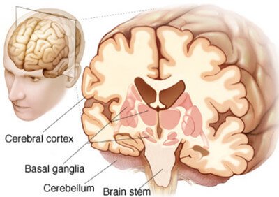
Picture 1: Structure of the Brain and Location of the Basal Ganglia
Image Source: neuroanatomyblog.tumblr.com

The Basal Ganglia image

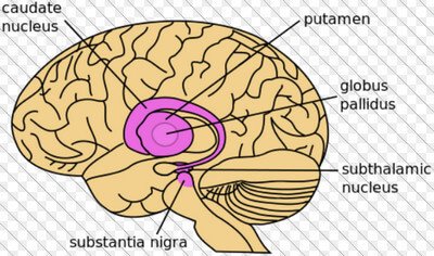
Picture 2: The Basal Ganglia
Image Source: upright-health.com

Parts of the Basal Ganglia photo

Picture 3: Parts of the Basal Ganglia
Image Source: commons.wikimedia.org

 Relationship of the parts of Basal Ganglia to the Thalamus and Ventricles

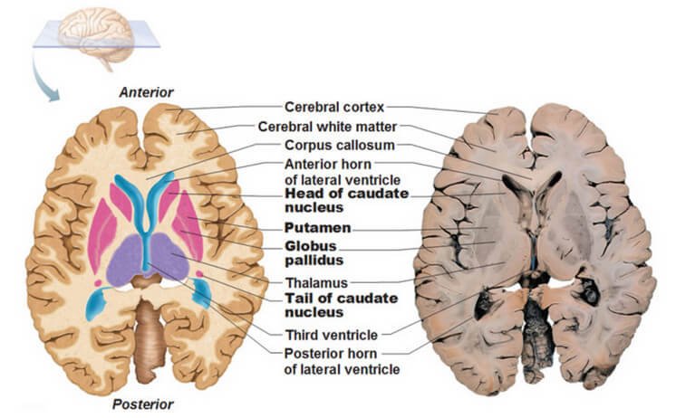
Picture 4: Relationship of the Basal Ganglia to the Thalamus and Ventricles
Parts of the basal ganglia are emphasized in bold letters.
Image Source: antranik.org

Blood Supply of the Basal Ganglia image

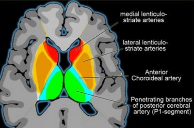
Picture 5: Blood Supply of the Basal Ganglia
Image Source: radiologyassistant.nl

What are the Functions of the Basal Ganglia?

  • The basal ganglia allow you to automatically perform a learned motor behavior.
  • From your motor memory, basal ganglia facilitates in preparing for motor action.
  • It controls and modifies your movements.
  • It is one of the brain structures that maintain posture.
  • Basal ganglia play a role in memory retrieval [2].

Stroke Definition

Medically termed cerebrovascular disease or cerebrovascular accident (CVA), stroke is referred to any pathologic disturbance in the blood vessels of the brain causing some parts of it to be deprived of blood and oxygen, resulting to neurologic deficits and possibly death. It may be ischemic or hemorrhagic [5].

Hemorrhagic Stroke vs. Ischemis Stroke photo

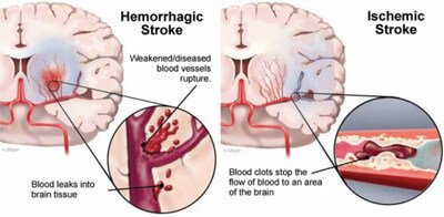
Picture 6: Hemorrhagic Stroke vs. Ischemis Stroke
Image Source: heart.arizona.edu

Risk Factors of Basal Ganglia Stroke

  • Hypertension occurs in 90% of the cases.
  • Moyamoya disease
  • Chronic alcoholism
  • Use of cocaine [3]

Signs and Symptoms of Bilateral or One-Sided Basal Ganglia Stroke

Cerebrovascular disease of the basal ganglia often shows motor dysfunctions. The severity of signs and symptoms depends on how extensive the damage is and which parts of the basal ganglia are specifically affected.
Limitation in Motor Activities

  • Ataxia or inability to coordinate muscles
  • Muscle weakness and rigidity
  • Involuntary tremors
  • Facial asymmetry
  • Pocketing happens when the mouth or throat is affected. This means the foods are only chewed or held on one side of the mouth.

Impaired Sensation

The patient will not be able to normally feel stimuli as he had before the stroke. He may not be able to feel touch, pain, temperature, or pressure in a certain area of his body. He may not even know which body part is being touched.
Speech Problems

  • Nonfluent Aphasia
    The patient has a problem with speaking his mind. What is in his mind is not completely what comes out of his mouth. There are missing words and incomplete sentences. He finds it difficult to speak.
  • Fluent Aphasia
    The patient does not find it hard to speak. He speaks fluently and in complete sentences. The problem here is the words itself. His sentences are jumbled words that have no meaning altogether. What the patient wants to say is completely different from what comes out of his mouth. The statements do not make sense at all.
  • Global Aphasia
    The patient cannot speak nor understand words.

Changes in the Eyes

  • Trouble looking upwards or sidewards
  • Loss of visual field in some areas
  • Pupils are asymmetrical in size

Personality Changes

  • Depression
  • Inappropriate affect
  • Inappropriate emotions
  • Rage
  • Frustration
  • Nervousness
  • Avolition or lack of motivation [6]

Right Basal Ganglia Stroke

  • Anosognosia is a state wherein the patient is not aware or unable to perceive the severity of his deficit. This is frequent among patients who had right-sided hemispheric stroke, affecting the right middle cerebral artery, which supplies parts of the basal ganglia.
  • Left-side neglect happens in patients who had basal ganglia stroke on the right side of his brain. The patient will unconsciously neglect or ignore anything that is on his left side. He only pays attention on what’s on the right side of his body. He may even have trouble moving his body parts to the left.
  • Infarction and haemorrhage of the right anterior choroidal and lenticulostriate arteries put the basal ganglia and internal capsule into the picture. There will be visuospatial hemineglect, constructional apraxia, motor impersistence, and anosognosia.
  • Infarction of right anterior choroidal artery does not cause impairment in memory [6, 7].

Left Basal Ganglia Stroke

  • Apathy, meaning lack of interest or concern as manifested by inactivity, occurs after the occurrence of lesion on the left basal ganglia.
  • Infarction of left anterior choroidal artery may cause impairment in memory.
  • Stroke in the left basal ganglia is associated with post-operative major depression. There is a study saying that major depression is twice likely to occur in left basal ganglia stroke than the right. It was also said that depressive disorder is more likely to occur in strokes located in the basal ganglia than in the thalamus, so as with middle cerebral artery than in posterior blood vessels [7, 8].

Neurologic Disorders associated with Basal Ganglia Stroke

Recurrent Artery of Heubner Syndrome

Recurrent artery of Heubner or medial striate artery is a branch of the anterior cerebral artery. Infarction of this blood vessel results to weakness of the face and arm contralateral to the affected side of the brain. Cognitive and behavioural abnormality may also be observed. The infarcted parts include the anterior part of the basal ganglia, inferior frontal lobe, and anterior internal capsule [2].

Thalamic Syndrome

A group of signs and symptoms including sensory loss, pain, dysesthesia, choreoathetosis, tremor, and hemiparesis occur if thalamogeniculate artery is occluded. Hemiballismus is an additional symptom if the subthalamic nucleus and globus pallidus are affected.

Transient Choreoathetosis

It comes from the root words “chorea” which is an uncontrollable, rapid movement and “athetosis” meaning twisting and wiggling movement of the hands and feet. During prolonged activity, choreoathetosis seems to trigger ischemia of the anterior basal ganglia [5].

Treatment for Basal Ganglia Stroke

Warning Signs of Stroke emergency

Picture 7:  Warning Signs of Stroke
Image Source: dhhs.ne.gov

Remember the image above. If there is someone with you and they manifest these signs, you better act fast and rush to the hospital as quick as you can. You may be saving that person’s life.

Once the patient gets into the emergency room, the physician provides measures to dissolve the clot and prevent further clot formation through blood thinning medications. Fluids are administered and vital signs are stabilized. Lowering the blood pressure and intracranial pressure is extremely important.

If not treated by medications, the patient undergoes into emergency surgery to remove the cause itself as it is fatal to the patient if the brain does not get enough blood and oxygen. Time is of the essence because the longer the brain becomes deprived, the higher the chance of disability and mortality.

After recovery, the patient gets into different kinds of therapies, depending on severity of damage the stroke. He signs into physical therapy, speech therapy, occupational therapy, and others which may help him regain his former functions. The goal here is to optimize the patient’s capabilities so he can live his life to the fullest.

It is also very important to prevent it from happening again by stabilizing the blood pressure and avoiding all the risk factors.

Prognosis of Basal Ganglia Stroke

  • Approximately 33% of all stroke cases are deadly. Prognosis depends on the underlying cause, how extensive it is, how soon it was medically treated, size and location of the lesion, degree of deficit, and age of the patient.
  • The chance of death for patients with hemorrhagic stroke is 70% while for ischemic stroke, mortality is lower which is 25%. However, reoccurrence of ischemic stroke is 5-15% every year [9].
  • Patients who had stroke confined to the basal ganglia have smaller lesions but slower initial recovery time compared to those who had stroke on the cerebral cortex. Although the recovery was gradual during early rehabilitation stage, it significantly progresses towards the end. Compared to patients who had stroke on cerebral cortex, those who had stroke on basal ganglia had a greater overall recovery [10].
  • The earlier the stroke was recognized and treated, the better the prognosis. The greater the Glasgow coma scale (GCS) score of the patient, the better the prognosis.

Prevention of Stroke

Any forms of CVA are dangerous to face. The best way to never experience stroke is prevention. In order to do that, risk factors (as enumerated in the earlier section of this article) to stroke should be eliminated.

The physician’s prudence in the identification of early signs of stroke such as transient ischemic attack (TIA), carotid artery stenosis, and atrial fibrillation is a very important element in saving a patient’s life [5].

References:

  1. Basal Ganglia accessed on http://en.wikipedia.org/wiki/Basal_ganglia
  2. Afifi AK & Bergman RA, Functional Neuroanatomy: Text and Atlas 2nd edition, McGraw-Hill 2005
  3. Caplan LR, Stroke Syndromes 3rd edition, Cambridge University Press, 2012, p 509
  4. Greenstein B & Greenstein A, Color Atlas of Neuroscience, Thieme 2000 , p 186
  5. Ropper AH & Samuels MA, Adams & Victor’s Principles of Neurology 9th edition, McGraw-Hill Companies Inc. 2009
  6. Basal Ganglia Stroke accessed on https://patienteducation.osumc.edu/Documents/BasalGangliaStroke.pdf
  7. Godefroy O, The Behavioral and Cognitive Neurology of Stroke, Cambridge University Press 2013, pp 37-38
  8. Schaller B, State-of-the-Art Imaging in Stroke, Nova Publishers 2007, p 80
  9. Lindsay KW et al, Neurology and Neurosurgery Illustrated 3rd edition, Churchill Livingstone 1997, pp 236-237
  10. Barnes MP et al, Recovery after Stroke, Cambridge University Press 2005, p 162

Xiphoid Process

$
0
0

Xiphoid Process and Pain Definition

Other names for the xiphoid process are processus xiphoideus, ensiform or xiphoid appendix. It rests at the 10th thoracic vertebra. It is the tiniest and lowest part of the sternum. Unlike the other two parts of the sternum namely manubrium and body, there are costal cartilages and ribs that are attached to the xiphoid process.

  • Attached to the anterior surface of the xiphoid process are the anterior costoxiphoid ligament and a section of the rectus abdominis muscle.
  • The posterior surface serves as an attachment to the posterior costoxiphoid ligament, transversus thoracis muscle, and diaphragm.
  • Xiphisternal joint connects the sternal body to the xiphoid process’ superior border.
  • There are articular facets on both sides of the superior border where the 7th ribs attach.
  • Abdominal aponeuroses are attached to the lateral borders of the xiphoid process.

The xiphoid process is made up of cartilage until the person reaches the age of 40 wherein it ossifies. In some cases, the xiphoid process fuses with the sternal body when the person reaches the old age. The ossification of the xiphoid process presents itself as a lump that is commonly thought by patients as a tumor that may cause cancer. They have to be reassured that it is a normal development of the xiphoid process [1, 2, 3].

Photos of Anatomy: Where is the Xiphoid Process Located?

Sternum is a part of the skeletal system picture

Picture 1: Sternum lies on the anterior midline aspect of the thoracic wall. There are three parts: manubrium (green), body (blue), and xiphoid process (violet).
Image Source: wikipedia.org

The xiphoid process is colored red image

Picture 2: The xiphoid process is colored red.
Image Source: wikipedia.org

Clinical Significance: What is the Function of Xiphoid Process?

The xiphoid process is very small anatomical structure. Despite of its size, it is very important because it serves as a landmark for:

  • Subcostal or infrasternal angle that is formed by the right and left subcostal margins and the xiphoid process
  • Lower margin of the heart
  • Superior end of the liver
  • Inferior limit of the anterior thoracic cage
  • Central tendon of the diaphragm [3]

Xiphoid Process Pain

Causes of Xiphoid Process Pain

Xiphoidalgia

Also known as xiphoidynia or hypersensitive xiphoid syndrome, xiphoidalgia presents as a deep sharp pain that is felt on the xiphoid process that worsens when pressed. Overeating and movement that involves the use of xiphoid process, such as bending, stooping, and twisting, aggravates the pain.

It resembles the chest pain in heart attack. The xiphisternal joint may appear swollen and feel sore. The pain may radiate to the back of the sternum, precordium, epigastrium, shoulders, and back. The patient suffers the pain for minutes to hours and may persist in weeks or months.

Patients with enlarged xiphoid process with xiphisternal angle of less than 160 degrees are more likely to develop xiphoidalgia. Though the pain may be too much, xiphoidalgia is self-limiting, meaning it goes away on its owneven without treatment [4, 5, 6].

CPR and Sternum Pain

The thoracic cage is designed to protect all of the surrounding structures from trauma or injury. But like anything else, it has a breaking point. If the sternum cannot handle any more pressure that comes to it, fracture and dislocation becomes the problem.

A common mistake in the performance of cardiopulmonary resuscitation (CPR) is the wrong placement of the rescuer’s hand on the patient’s chest. When it is too low, xiphoid process may be fractured and protrude on the underlying organs. A sign when it becomes broken is when you sense a “pop”  [7, 8].

Also see : Pain under right rib cage

Gastroesophageal Reflux Disease (GERD)

GERD refers to the regurgitation of gastric acid from the stomach to the esophagus.

The esophagus is an alkaline environment, which is the total opposite of the acidic environment of the stomach. It is located behind the sternum. The esophagus runs from the throat down to the stomach. Anatomically speaking, the proximity of the esophagus and xiphoid process, explains why you feel sternum pain in GERD.

Moreover, the trigger points for somatovisceral symptoms such as heartburn lies surrounding the xiphoid process that is why you feel xiphoid process pain in GERD [9].

Treatment for Xiphoid Process Pain

  • Analgesics and nonsteroidal anti-inflammatory drugs (NSAIDs) are the first line of painkillers that may be given to patients with xiphoid process pain.
  • If it cannot be handled by the first line agents, opioid analgesics may be necessary to decrease the pain.
  • Injection of lidocaine or corticosteroids into the xiphisternal joint is eventually administered if none of the above works.
  • Heat application over the xiphoid process provides relief.
  • Gentle exercise of chest wall muscles gives you a better chance of mobility in the long run.
  • Elastic rib belt provides pressure to the sternal area thereby diminishing the pain [5, 6].

References:

  1. Gray H, Anatomy of the Human Body, Bartleby Bookstore
  2. Moore KL et al, Clinically Oriented Anatomy 6th edition, Lippincott Williams & Wilkins 2010, pp 76-78
  3. Snell RS, Clinical Anatomy by Regions 9th edition, Lippincott Williams & Wilkins 2012, p 35
  4. Fishman SM, Bonica’s Management of Pain, Lippincott Williams & Wilkins 2012, p 1063
  5. Klippel JH et al, Primer on the Rheumatic Diseases, Springer Science and Business Media 2008, p 84
  6. Waldman SD, Atlas of Uncommon Pain Syndromes, Elsevier Health Sciences 2013, p 193-194
  7. Tortora GJ & Derrickson B, Principles of Anatomy & Physiology 13th edition, Biological Science Textbooks Inc. 2012, p 245
  8. Ellis H, Clinical Anatomy 11th edition, Blackwell Publishing 2006, p 11
  9. Ferguson LW & Gerwin R, Clinical Mastery in the Treatment of Myofascial Pain, Lippincott Williams & Wilkins 2005, p 316

Endosteum

$
0
0

Endosteum : Definition and Functions

The endosteum is a structure in the middle of bone tissue and bone marrow. It is a thin covering that surrounds the medullary cavity. It coats the inner compact bone and the trabeculae of the spongy bone. It covers the loose structures found inside the bone. It is made up of connective tissue and one layer of cells.

The osteogenic capability of the endosteum lies on the fact that it houses osteoprogenitor cells or preosteoblasts that differentiate into osteoblasts (bone-forming cells), bone matrix-secreting cells, or bone-lining cells.

Osteoprogenitor cells are often called endosteal cells. It is hard to recognize even under the microscope. It is described to be flat with elongated nucleus. The features of the cytoplasm are vague.

Together with the periosteum, endosteum is responsible for growth of the bone in diameter but contrary to the periosteum, the fibrous layer of the endosteum is indistinct [15].

Photos of Anatomy and Histology


Periosteum vs. Endosteum picture

Picture 1: Periosteum vs. Endosteum
Image Source: wikipedia.org

 Skeletal tissues showing how endosteum lines the body canals and covers trabeculae image

Picture 2: Skeletal tissues showing how endosteum lines the body canals and covers trabeculae
Image Source: classes.midlandstech.edu

 Parts of the Skeletal Tissue photo

Picture 3: Parts of the Skeletal Tissue
Image Source: rci.rutgers.edu

 Endosteum and Other Parts of the Bone picture

Picture 4: Endosteum and Other Parts of the Bone
Image Source: knowyourbody.net

Endosteum on Cross-Section image

Picture 5: Endosteum on Cross-Section
Image Source: mhhe.com

Histology of the bone showing periosteum and endosteum photo

Picture 6: Histology of the bone showing periosteum and endosteum
Image Source: bvetmed1.blogspot.com


Clinical Importance

Chemical exchange between the bone marrow and blood vessels becomes possible through the osteoblasts of the endosteum.

Endosteal cells are active in the process of bone growth, repair, and remodelling. There are parts of the bone matrix that are not entirely covered by the endosteum. These exposed areas are the attachment sites for osteoblasts and osteoclasts wherein they can deposit or remove components of the bone matrix.

Endosteum and periosteum contribute to bone repair and reconstruction after a fracture occurs.  Blood vessels and tissue surrounding the injured area bleed and eventually form a clot through the edges of the broken bone.

The cells of endosteum and periosteum undergo rapid mitosis and proliferation then they migrate towards the affected site. There will be formation of a new bone that will serve as a bridge which temporarily stabilizes the broken ends of the bone [6, 7].

Endosteal Scalloping picture

Picture 7: Endosteal Scalloping
Image Source: radiopaedia.org

If medullary lesions develop along the inner aspect of the cortical bones, especially in the long bones, endosteal scalloping may be observed. Although these medullary lesions may be slow-growing and non-infiltrating, endosteal scalloping is associated with both benign and malignant conditions which include enchondroma, osteomyelitis, chondromyxoid fibroma, skeletal amyloidosis, periprosthetic osteolysis, brown tumor, chondrosarcoma, multiple myeloma, and skeletal metastasis [8].

In nuclear medicine radiation dosimetry, a normal endosteum measures 10 µm in thickness. Endosteal thickness of 50 µm from the medullary cavity and trabeculae suggest sensitivity of radioactive cells which makes a person prone to bone cancer [9].

Endosteum vs. Periosteum

Periosteum and endosteum are both parts of the bone. Aside from their rhyming words, many are still puzzled with the difference of these two. Here is a table summarizing these two very important parts of the bone [10].
Table 1: Comparison between Endosteum and Periosteum

References

  1. Ross MH & Pawlina W, Histology: A Text and Atlas 6th edition, Lippincott Williams & Wilkins, 2011, p 221
  2. Tortora GJ & Derrickson B, Principles of Anatomy and Physiology 13th edition, Biological Science Textbooks Inc., 2012, p 184
  3. Johnson KE, Histology and Cell Biology 2nd edition, Williams & Wilkins, 1991, pp 112-114
  4. Eroschenko VP, diFiore’s Atlas of Histology with Functional Correlations 11th edition, Lippincott Williams & Wilkins, 2008, p 90
  5. Henrikson RC & Mazurkiewicz JE, Histology Volume 518, Lippincott Williams & Wilkins, 1997, p 131
  6. Khan R, A Textbook of Biotechnoloy Volume 2, Firewall Media, 2007, pp 62-65
  7. Martini F et al, Anatomy and Physiology, Rex Bookstore Inc., 2007, pp 135 & 144
  8. http://www.ehealthstar.com/anatomy/endosteum
  9. McParland BJ, Nuclear Medicine Radiation Dosimetry, Springer Science and Business Media, 2010, p 512
  10. Weerakkody Y & Gaillard F, Endosteal Scalloping accessed on http://radiopaedia.org/articles/endosteal-scalloping


Patella Alta

$
0
0

Patella Alta : Definition

Patella alta or high-riding patella is an anatomical variation and bone malalignment of the patella (kneecap) in relation to the surrounding bones. The patella is structurally placed higher in the femoral trochlea than it should be.
Patella baja or infera is the opposite of patella alta. It is a low-riding patella that is structurally placed lower than it should be.

Causes

Patella alta is not hereditary. It may be caused by overgrowth of the patellar tendon during puberty, resulting to a high-riding patella.

The lateral femoral sulcus prevents lateral patellar subluxation. If the patient has patella alta, this protective feature of the lateral femoral sulcus will not be used [1].

Patella alta may be associated with the following conditions [2]

  • Patellar subluxation (recurrent)
  • Patellar dislocation
  • Chondromalacia of the patella
  • Patellar ligament rupture
  • Femoral osteomyelitis
  • Poliomyelitis
  • Cerebral palsy
  • Sinding-Larsen-Johansson disease (avascular necrosis of an ossification center of the patella)
  • Osgood-Schlatter disease (avascular necrosis of the tibial tuberosity)

 Lateral knee radiography shows complete tear of the tendon in traumatic patella alta picture

Picture 1: Lateral knee radiography shows complete tear of the tendon in traumatic patella alta.
Image Source: Scuderi GR

 Patella alta in cerebral palsy features a lengthened and high positioned patella due to chronic spasms image

Picture 2: Patella alta in cerebral palsy features a lengthened and high positioned patella due to chronic spasms.
Image Source: Scuderi GR

 Patella alta and prepatellar bursitis can be observed in patients with cerebral palsy due to crawling photo

Picture 3: Patella alta and prepatellar bursitis can be observed in patients with cerebral palsy due to crawling.
Image Source: Scuderi GR

Measurement and Diagnosis


Camel back Sign picture

Picture 4: Camel back Sign
Image Source: Scuderi GR, The Patella, Springer Science and Business Media, 1995, p 73

With the patient in standing position, the examiner goes to the patient’s side and assesses for the presence of camel back sign of the knee.

Camel back sign is observed in patients with patella alta. The first hump is the high positioned patella and the second hump is represented by the infrapatellar fat pad or infrapatellar bursa.

Grasshopper Eyes image

Picture 5: Grasshopper Eyes
Image Source: Scuderi GR, The Patella, Springer Science and Business Media, 1995, p 74


Normally, patellae should be directed towards the front when in a sitting position. In patella alta, patellae are directed upwards.

Grasshopper eyes or frog eyes is seen in patients whose patellae are proximally displaced (patella alta) and externally or laterally rotated (lateral patellar tilt) [3, 4].

a) Blumensaat's Line; b) Insall-Salvati Measurement; c) Blackburn-Peel Measurement photo

Picture 6: a) Blumensaat’s Line; b) Insall-Salvati Measurement; c) Blackburn-Peel Measurement
Image Source: Paley D, Principles of Deformity Correction, Springer Science and Business Media, 2002, p 569

Blumensaat’s line is an imaginary line formed with 30° flexion of the knee. The inferior border of the patella should be in line with the extension of intercondylar notch.

In Blackburn-Peel measurement, the quotient of (i) tibial plateau to the lower end of the patellar articular surface and (ii) patellar articular surface length is 0.8. If the quotient is greater than 1, the patient has patella alta [5].

In Insall-Salvati measurement, a lateral radiograph of the knee in 30° flexion is obtained. The ratio for the length of patellar tendon (LT) versus length of the patella (LP) is 1, meaning they should be equal. ±0.2 indicates an anatomical variation.

A ratio of 1.2 means LT is longer than LP, which makes the patella located in a higher position than it should be. This indicates patella alta. On the other hand, a ratio of 0.8 indicates patella baja, meaning the LT is short, which makes the patella located in a lower position [1, 6].

A study in Boston University (2010) revealed that a high Insall-Salvati ratio increases the risk of having cartilage damage, bone attrition, and bone marrow lesions. Patella alta and lateral trochlear inclination could be factors that contribute to the development of patellofemoral joint osteoarthritis [7].

Measuring the LT is important because if it is increased, as in the case of patella alta, more flexion than the usual should be performed in order to allow the patella to rest on the trochlea.
Treatment

Persons with patella alta may have patellar pain and gait instability. This warrants the patient to submit himself into treatment or therapy. The goal of treatment is to resume the normal function of the knee and to relieve any forms of discomfort.
Conservative Treatment

Conservative treatment for problems in patella may respond well and may possibly get surgery out of your list of options. Patella alta may be corrected through [8]:

  • Manual gliding
  • Physical therapy including closed chain strengthening and exercise programs
  • Patellar taping
  • Braces or sleeves for knees
  • Pain relievers
  • Ice packs
  • Weight loss

Surgery

Tibial tuberosity osteotomy can be performed in patients with patella alta. This solves the problem by moving the attachment of the patellar ligament to the tibia downwards. That way, patella goes down with it.

If an increased quadriceps (Q) angle comes with patella alta, tibial tuberosity osteotomy can correct it too by shifting the bony attachment of the patellar ligament inwards.

Complications of surgery may include infection, stiffness of knee joints, deep vein thrombosis (DVT), nerve injury, and recurrent instability.

Knee splint is applied after surgery and stays with the patient for 6 weeks. The patient is expected to use crutches. Knee movement will be restricted. Weight bearing is limited and is only allowed with the knees straight. Physical therapy and exercise is required after surgery in order to get rid of the pain and swelling and resume usual muscle control. This will also prevent further complications. Recovery period is around 3-6 months [9].

References:

  1. Hammer WI, Functional Soft-tissue Examination and Treatment by Manual Methods, Jones & Bartlett Learning, 2007, pp 328-329
  2. Burgener FA & Kormano M, Differential Diagnosis in Conventional Radiology, Thieme, 2011, p 317
  3. Scuderi GR, The Patella, Springer Science and Business Media, 1995
  4. Magee DJ, Orthopedic Physical Assessment, Elsevier Health Sciences, 2008, p 737
  5. Paley D, Principles of Deformity Correction, Springer Science and Business Media, 2002, p 569
  6. Baratz M et al, Orthopedic Surgery: The Essentials, Thieme, 1999, p 544
  7. Stefanik JJ, Patella Alta and Its Relationship with Patellofemoral Joint Alignment and Osteoarthritis, Boston University, 2010
  8. http://www.genufix.com/patella_problems1.htm
  9. http://www.kneesurgeon.com.au/patella-trochlear-dysplasia.html

Shoulder Blade Pain

$
0
0

Shoulder blades or scapulae are the triangular shaped bones located on your upper back below and behind the shoulders. Right and left shoulder blades serve as the anchor for muscles that contribute to movements of the upper trunk, shoulders, and arms. Its function in everyday movements makes it difficult to ignore the pain felt in, under, behind, below, around, or near the shoulder blades.

Usually, the pain goes away with rest along with other home remedies. If severe pain happens suddenly that is not preceded by any activity or if the pain gets worse as the days go by, it may be necessary for you to visit your doctor.

General Causes of Shoulder Blade Pain

  • Muscle Strain: Any overused and abused muscle causes muscle strain. Sleeping in an uncomfortable position especially at night is one of the most common reasons why a person experiences pain around the shoulder blades when he wakes up. Muscle strain also happens when you carry or lift heavy objects when you are not used to do so. Strenuous exercise or sports such as tennis or golf may develop shoulder blade pain.
  • Rotator Cuff Injury: The rotator cuff consists of four muscles and tendons that stabilize the shoulder. If one of the supraspinatus, infraspinatus, teres minor, and subscapularis muscles is injured, there will be pain of the shoulder blade in the affected side. This injury is common to athletes.
  • Fracture: Scapulae are rarely fractured but it could happen due to vehicular accident, sports injury, or fall.
  • Osteoporosis and Arthritis: Porous bones and inflammation of the joints around the shoulder blades, shoulders, or neck may cause pain around the shoulder blades.

Shoulder Blade Pain

  • Nerve Impingement: Burning pain on the shoulder and/or shoulder blade that radiate to your arm may signify an impinged or damaged nerve. Your fingers may perceive burning sensations as well. It may last for weeks or months. This does not go away on its own. It needs medical attention.
  • Lung Tumor: Tumors in the lung, especially pancoast tumor or the one that lies on superior aspect of the lung, may cause pain behind the shoulder blades. Pneumothorax and pulmonary embolus may also cause shoulder blade pain.
  • Herniated Disk: Presence of slipped disks in the neck region causes referred pain to the shoulder blades.
  • Varicella Zoster Virus: This virus that causes chickenpox and shingles may cause shoulder blade pain. In shingles, one of the early symptoms is burning pain on the right shoulder blade.
  • Cancer: When a cancer, whichever the source is, metastases to the bones, pain may be felt around the shoulder blades [1, 2].

Left Shoulder Blade Pain

Left shoulder blade pain in women may be indicative of myocardial infarction (MI) or heart attack. Along with this, the patient also complains of pain in the chest, jaw, and upper back.

Other signs and symptoms include feelings of indigestion, fatigue, palpitations, impending doom, pallor, cold clammy skin, difficulty of breathing, excessive sweating, hypotension or hypertension, anxiety, restlessness, crackles, heart murmurs, and atrial gallop. Pain in the right shoulder blade seldom occurs with MI [3].

Inflammatory disorders such as pericarditis, pancreatitis, and pleuritis cause referred pain to the left shoulder blade. It can also be a symptom for other health conditions such as aortic dissection, pulmonary embolism, and pneumonia [4].

Right Shoulder Blade Pain

Diseases of the gallbladder are manifested by a referred pain to the right shoulder blade. Fat women in their 40s are at risk in developing problems with the gallbladder.

The gallbladder is an organ located at the right upper quadrant below the liver. It stores bile that is responsible for chemically digesting fats or lipids. Everytime you eat fatty foods, your gallbladder becomes active as it secretes the bile.

If your gallbladder is diseased or has gallstones in it, you will experience severe sharp pain after eating. The pain radiates up to your right shoulder blade.

Different diseases of the liver may also cause pain to the right shoulder blade. An infected liver creates pus. Liver cirrhosis leads to peritonitis or fluid accumulation in the abdomen. Liver tumor or cancer may metastasize to different parts of the body. All of these result to general feelings of illness and pain behind the right shoulder blade [5].

Pain between the Shoulder Blades

Upper back pain between the shoulder blades is connected with the Causes section of this article. Generally, it may be due to muscle strain, rotator cuff injury, fracture, osteoporosis, arthritis, nerve impingement, herniated disk, virus, or cancer.

If you have a problem with your ribs, stabbing pain when breathing may be felt. Sharp pain after eating is felt when you have a problem with your gallbladder.

Severe pain between shoulder blades may be associated with thoracic outlet syndrome, rib problem, lung process, or pathologic diseases of the thoracic spine.

Diagnosis

  • The doctor begins by taking comprehensive history and physical examination. However, this may not be enough to make a reliable diagnosis.
  • Radiography including, chest x-ray, MRI, CT scan, or PET scan may be necessary to aid the diagnosis.
  • If a heart disease is suspected, the doctor will order you to have ECG and stress test.
  • Endoscopy or other abdominal exams is performed to determine if cause of shoulder blade pain is a problem in the gastrointestinal system [1].

Treatment for Shoulder Blade Pain

Shoulder blade pain has many causes. Treatment depends on it [2].

  • If it is as simple as a muscle strain, it’s easy. Do your shoulder blades a favour and take a rest.
  • Maintain proper posture and biomechanics.
  • Heat and cold compress offers relief.
  • Massage provides comfort.
  • At night, sleep in a comfortable position with your head and shoulders aligned so you won’t have to wake up in the morning having a sore shoulder blade. Use pillows.
  • Perform scapular retraction exercises. Position your arms at shoulder level and imagine that someone is standing right in front of you. Wrap our arms around that imaginary person and hold for 8-10 seconds.
  • If the pain is severe, muscle relaxants and even steroids are prescribed by the doctor.
  • If the cause is cancer, the treatment can get as complex as chemotherapy, radiation therapy, and surgery.

References:

  1. Eldridge L, Shoulder Blade Pain, About.com, 06/20/14 accessed on http://lungcancer.about.com/od/Symptoms-Of-Cancer/a/Shoulder-Blade-Pain.htm
  2. http://www.md-health.com/Shoulder-Blade-Pain.html
  3. Interpreting Signs and Symptom, Lippincott Williams & Wilkins, 2007, p 129
  4. http://www.buzzle.com/articles/pain-under-left-shoulder-blade.html
  5. http://www.md-health.com/Right-Shoulder-Blade-Pain.html

Lingual Tonsils

$
0
0

Definition and Function

Lingual tonsils are a group of lymphoid nodules (30-100 follicles) located at the posterior third of the tongue.

Their appearance is described as exophytic mucosal small masses with crypts and folds. This creates the reticular pattern at the base of the tongue. Its precise location is between the circumvallate papillae and the lining of vallecula that extends to the root of epiglottis [1, 2].

The tonsils are parts of the lymphatic system that wards off harmful substances and infection. The lingual tonsil basically protects us from upper respiratory tract infections. The lingual tonsil enlarges from birth until the person reaches 7 years of age and eventually shrinks as one grows old [3].

Anatomy and Histology

The Waldeyer’s ring consists of the pharyngeal, palatine, and lingual tonsils picture
Picture 1: The Waldeyer’s ring consists of the pharyngeal, palatine, and lingual tonsils.
Image Source: smilingldsgirl.com

The Dorsum of the Tongue image

Picture 2: The Dorsum of the Tongue
Image Source: en.wikipedia.org

Sagittal Section of the Oral Cavity photo

Picture 3: Sagittal Section of the Oral Cavity
Image Source: studyblue.com

Lingual Tonsil Histology picture

Picture 4: Lingual Tonsil Histology
Image Source: cea1.com

Lingual Tonsil Histology image

Picture 5: Lingual Tonsil Histology
Image Source: php.med.unsw.edu.au

The lingual tonsil is lined by a nonkeratinizing squamous epithelium. A definitive histological feature of a tonsil is the presence of tonsillar crypts. These start to appear after birth. Lingual tonsils have shallower crypts and appear less branched when compared to palatine tonsils.

Other important structures observed under the microscope are the lymphoid follicles, mucous salivary glands, skeletal muscles, and adipose tissue.

The lymphatic vessels of the posterior portion of the tongue pass through the pharyngeal wall around the external carotid artery and drain into the deep cervical lymph nodes [4].

Clinical Correlation

Enlarged and Swollen Lingual Tonsil

The lingual tonsil becomes enlarged and painful in the presence of inflammation, allergy, or infection. It can also happen as a compensatory mechanism after the patient has undergone tonsillectomy or adenoidectomy.

Normally, the lingual tonsils have a reticular pattern on its surface. With hypertrophy of the lingual tonsils, this pattern will be disturbed.

Frontal view of the radiograph will show 5-7 mm, smooth surfaced nodules in patients with lymphoid hyperplasia. Laterally, these nodules distend posteriorly. In severe cases, these nodules reach the vallecula.

Unilateral enlargement of the lingual tonsil warrants tissue biopsy in order to rule out lymphoma or squamous cell carcinoma [1, 2]. Lymphoma increases in size and is more likely to obstruct the airway. Squamous cell carcinoma, on the other hand, is the more common type of cancer of the lingual tonsil. It appears as ulcers at the back of the throat [5].

Lingual Tonsil Obstructing the Airway photo

Picture 6: Lingual Tonsil Obstructing the Airway
Image Source: Kummer A, Cleft Palate & Craniofacial Anomalies: Effects on Speech and Resonance, Cengage Learning, 2007

The lingual tonsil is located at the back of the tongue, meaning it is close to the airway. With its enlargement, it may cause airway obstruction and breathing difficulties. It may come to a point where the larynx cannot be seen and the vallecula may be covered with tissues of the lingual tonsil, as illustrated above [6].

Lingual Tonsillitis

Lingual Tonsillitis

Picture : Lingual Tonsillitis
Image Source: simple-health-secrets.com

Lingual tonsillitis is primarily presented as a complaint of pain over the hyoid bone, which may be unilateral or bilateral. Upon physical examination, the lingual tonsils appear swollen and exudates are noted to be present. Other signs and symptoms include fever, dysphagia (difficulty in swallowing), tenderness over the affected area, lymphadenopathy, and muffled voice [7].

More often than not, bacteria is the cause of lingual tonsillitis so preliminary antibiotics is being prescribed. Throat swab is obtained and cultured. If bacteria is ruled out to be the cause, antibiotics will be discontinued. If lingual tonsillitis is caused by a virus, symptomatic treatment will be ensued. Pain medications will be prescribed. Gargling with saline solution seems to work well [5].

Oral Lymphoepithelial Cyst or Cystic Ectopic Lymphoid TissuePicture  : Oral Lymphoepithelial Cyst or Cystic Ectopic Lymphoid Tissue
Image Source: intechopen.com

The most common locations of oral lymphoepithelial cysts or cystic ectopic lymphoid tissue are the lateral sides of lingual tonsils, floor of the mouth, and tongue surface.

This occurs when the tonsillar crypts become housed with keratin and this will form the cysts. They appear as whitish or reddish yellow dome-shaped nodules. These cysts will enlarge due to inflammatory or allergic reactions. Airway obstruction warrants its removal [8].

Lingual Tonsillectomy

Indications for lingual tonsillectomy include its enlargement causing airway obstruction and obstructive sleep apnea, chronic and recurrent infections, and cancer or malignancy.

Under general anesthesia, the tongue is pulled out with the lingual tonsils clamped. Preferred removal of the lingual tonsil is through laser or cautery in order to minimize bleeding.

Watch out for postoperative hemorrhage. Aspirin is contraindicated post-tonsillectomy to avoid bleeding.
Postoperative pain is most intense for the first two days then it wanes off. Four to six days after surgery, the pain comes back and this time, the pain radiates to the ears. Scabs will develop at the back of the throat and this may cause halitosis (bad breath).

After the surgery, pain medications are prescribed in order to relieve the discomfort. Antibiotics are given for halitosis and to prevent postoperative infection. These will be taken in for 10 days. Ice chips or popsicles may help ease the swelling.

Typically, the post-operative patient may resume clear fluid diet followed by soft diet then diet as tolerated. The patient should not eat hot, spicy, acidic, sour, rough, and scratchy foods because these will add up to the already agonizing pain.

If the lingual tonsil is verified as cancerous or malignant, chemotherapy, radiation therapy, and another surgery may be necessary [9].

References:

  1. Ekberg O & Aksglaede K, Radiology of the Pharynx and the Esophagus, Springer Science & Business Media, 2004, pp 60-61
  2. Bruch JM & Treister N, Clinical Oral Medicine and Pathology, Springer Science & Business Media, 2009, pp 5 & 19
  3. http://www.innerbody.com/anatomy/nervous/lingual-tonsil-side#full-description
  4. Cardesa A & Slootweg P, Pathology of the Head and Neck, Springer Science & Business Media, 2006, p 184
  5. http://www.wisegeek.org/what-is-a-lingual-tonsil.htm
  6. Kummer A, Cleft Palate & Craniofacial Anomalies: Effects on Speech and Resonance, Cengage Learning, 2007, p 288
  7. Interpreting Signs & Symptoms, Lippincott Williams & Wilkins, 2008, p 591
  8. Burket LW, Burket’s Oral Medicine, PMPH-USA, 2003, p 114
  9. http://advancedotolaryngology.com/patient-education/tonsillectomy/

Mastoid Process

$
0
0

What is the Mastoid Process and Function

The mastoid part of the temporal bone houses the mastoid process. Mastoid process is the bony prominence easily felt behind the earlobe. It is one of the key features of the lateral cranium. It is located behind and below the external auditory meatus. It primarily functions as attachment to the neck muscles.

Inside of it are mastoid air cells or sinuses that are prone to inflammation and infection. These are cells are connected to the middle ear. Mastoid air cells are covered by mucoperitoneum that is continuous with the tympanic cavity and with the squamous part of the temporal bone [1].

The mastoid notch is located medial to the mastoid process. It is pierced by stylomastoid foramen in front and mastoid foramen is found on its back. Mastoid notch serves as the site of muscle attachment for the anterior and posterior bellies of the digastrics whose function is to open the mouth [2].

At birth, mastoid process may not be palpated. Its protrusion is due to the pulling of sternocleidomastoid muscle of the neck when a person starts to move his head. It only appears as a bony projection once the child turns 2 [3].

Where is the mastoid process ?

Photos of Anatomy and Location
Location of Mastoid Process picture

Picture 1: Site of Mastoid Process
Image Source: pennmedicine.org

Lateral view of the skull highlighting the mastoid process image
Picture 2 Lateral view of the skull highlighting the mastoid process.
Image Source: studyblue.com

Inferior view of the skull highlighting the mastoid process photo
Picture 3 : Inferior view of the skull highlighting the mastoid process.
Image Source: studyblue.com

Posterior View of the Skull showing the Mastoid Process picture

Picture 4: Posterior View of the Skull showing the Mastoid Process
Image Source: gl1800riders.com

Mastoid Cells of Lenoir image

Picture 5: Mastoid Cells of Lenoir
Image Source: wikipedia.org

Mastoid Process and its Relationship with the Middle Ear photo

Picture 6: Mastoid Process and its Relationship with the Middle Ear
Image Source: studyblue.com

Clinical Importance

No Mastoid Process in Newborns
Lateral skull of a newborn shows no mastoid process picture

Picture 7: Lateral skull of a newborn shows no mastoid process.
Image Source: cnx.org

A newborn has no mastoid process. This means that there is less protection for the facial nerve or CN VII after birth. This nerve arises from the stylomastoid foramina and since there is no mastoid process yet, it develops close to the surface. This makes it prone to injury or damage during surgical procedures such as forceps delivery or operations for treating middle ear problems.

The mastoid process starts to develop after the patient turns 1 year old. That is when the sternocleidomastoid muscles pull on the petromastoid parts of the temporal bones. Mastoid process is already developed by the age of 2 [4].

How Infection Spreads from the Middle Ear to the Brain

Mastoid Air Cells and the Nearby Structures image

Picture 8 Mastoid Air Cells and the Nearby Structures
Image Source: teachmeanatomy.info

Note the close proximity of the abscessed mastoid antrum to the sigmoid sinus and cerebellum photo

Picture 9: Note the close proximity of the abscessed mastoid antrum to the sigmoid sinus and cerebellum.
Image Source: smokh.org

The mastoid antrum is a small cavity found at the back of the petrous part of temporal bone. By way of aditus, it serves as a bridge between the mastoid air cells, posterior wall of the middle ear, and the sigmoid sinus and cerebellum of the brain. It is through the mastoid antrum that infection is spread from the ears to the brain [1].

Mastoiditis

Mastoiditis is caused by bacterial infection causing the ear, particularly the posterior area, to appear swollen and picture

Picture 10: Mastoiditis is caused by bacterial infection causing the ear, particularly the posterior area, to appear swollen and enlarged.
Image Source: fairview.org

Mastoiditis is the inflammation and infection of the mastoid bone. This disease is common among children and was once known as one of the most common cause of mortality among them primarily because medications find it hard to reach its target, which is the mastoid bone. Furthermore, the infection could spread to the brain.

Mastoiditis is caused by ear infections, particularly by otitis media. As explained earlier, the mastoid air cells are connected to the middle ear. It can also be caused by cholesteatoma, which will be discussed further in the next sections of this article.

Signs and symptoms of mastoiditis include ear pain, ear drainage, swelling and redness around the ear, enlarged ear, impaired hearing, fever, and headache.

Treatments for mastoiditis include antibiotics, ear drops, and ear cleaning. Myringotomy (fluid drainage of the middle ear) and mastoidectomy (removal of the infected mastoid bone) are options if the disease is severe.

Mastoiditis should not be left untreated because it in the long run, it might result to hearing loss, meningitis, or brain abscess that can lead to death [5, 6].

Parotiditis and Mumps

Note the proximity of the infected site to the mastoid process photo

Picture 11: Note the proximity of the infected site to the mastoid process.
Image Source: healthimpactnews.com

Inflammation and infection of the parotid gland causes severe pain that is aggravated when chewing. The parotid sheath that surrounds the parotid gland is tough and when the gland swells, the sheath limits its swelling, thereby producing severe pain.

The mastoid process comes into the picture when the pain is aggravated when opening the mouth and chewing. As the mouth opens, the posterior border of the mandibular ramus moves downwards and backwards, compressing the mastoid process. This is the reason why it hurts to eat when you have parotiditis or mumps [4].

Cholesteatoma


Cholesteatoma picture
Picture 12: Cholesteatoma
Image Source: chroniclescamera.blogspot.com

Cholesteatoma is an ear disease wherein a benign skin cyst pervades the mastoid process and the middle ear. This occurs when the Eustachian tube, which normally equalizes the ear pressures, does not open enough to perform its function. As a result, the tympanic membrane or eardrum becomes perforated and retracted as it forms a pocket that accepts the skin cyst.

The patient with cholesteatoma has a history of chronic ear infections. Alongside with this, there is ear discharge and gradual hearing loss.

Cholesteatoma may be treated through surgical procedures such as tympanoplasty, mastoidectomy, or ossiculoplasty [6].

Mastoid Cancer

Malignant lumps and tumors of the mastoid are mostly due to squamous cell carcinoma. It is a form of skin cancer. People who have chronic mastoid infection, family history of skin cancer, and exposure to coals, tars and arsenics are more prone to develop mastoid cancer [6].

References:

  1. Ellis H, Clinical Anatomy 11th edition, Blackwell Publishing Ltd., 2006, p 386
  2. Saladin K, Anatomy & Physiology: The Unity of Form and Function 5th edition, The McGraw-Hill Companies Inc., 2009, p 252
  3. Snell RS, Clinical Anatomy by Regions 9th edition, Lippincott Williams & Wilkins, 2012, p 663
  4. Moore KL et al, Clinically Oriented Anatomy 6th edition, Lippincott Williams & Wilkins, 2010, pp 840 926
  5. http://www.webmd.com/cold-and-flu/ear-infection/mastoiditis-symptoms-causes-treatments
  6. http://www.livestrong.com/article/190151-diseases-of-the-mastoid/

Kidney Pain

$
0
0

Known mostly for their waste removing functions in our bodies, kidneys are bean shaped organs. They look like two sprouting beans attached opposite each other and separated by venacava, the aorta, and other gastrointestinal organs.

Kidney Location (Anatomy)

  • Anatomically, they are positioned on your back; or more precisely,
  • Just under both the lower ribs (4 ).
  • The areas may probably and clearly explained as the upper abdominal area but on the backside usually near the back muscles.
  • They are intricately hidden in the abdominal region, right next to the liver and the spleen
  • Owing to their peculiar opposite positions, both kidneys are on not the same level.
  • The right kidney lies lower and just under the diaphragm, while the left kidney being slightly higher due to the large size of the liver.

Kidney location

Picture 1 : Where are the kidneys located in relation to quadrants?

The delicate and vital organs, are surrounded by layers of fat, and are protected by the ribcage, muscles, as well as each having a layer of connective tissues. On the back, they will feel as though they are deeply embedded on each side of the spine as depicted in the diagram below (1).

On a surface, here is where the Kidney is found

kidney surface anatomy


kidney surface anatomy back


Viewed from behind, the kidneys shown by the arrow and appear right at the inner part of the lower ribcage area. The left kidney is positioned higher on the ribcage area and next to the spleen. It may appear smaller in size in women and children as opposed to the male gender. The right kidney slighly lower is is not tottally enclosed by the ribcage and liver and the lungs.

position of each kidney viewed from the front side

The above diagram shows the position of each kidney viewed from the front side.

Various Types of Kidney Pain

There are several types of pains attributed to the kidney, most of which are in the back area of your body and often in the upper region near the ribcage areas, just above the hips. Unlike the lower back pains that are most severe (5);

  • Most kidney pains will be dull
  • Felt on either side of the spine region.
  • However, the pain can be quite painful especially when there is presence of kidney lacerations or kidney stones.
  • One paramount difference with other back pains is that all kidney pains will be accompanied by high fevers, nausea and in some cases lead to vomiting.
  • The pains caused by kidney stones will be wavy-like, moving from the upper backside to the front hip area (2).
  • Unlike back pains that will restrict back movements, severe kidney pains are comparable to Labour pains, affecting the movement of every other part of the body.
  • With back pains, one feels the pain on simple movements of the body; it actually becomes difficult to move. Kidney pains will be delayed after a move although in severe cases, the pain may be more elaborated and affect the body movements or exercises.

How to know if you are experiencing Kidney Pain

  • Most kidney pains will be dull. This is especially when kidney disease is on the onset.
  • With chronic kidney disease, the pain will however be severe.
  •  Since onset kidney pains are rarely specific, they can be confused with side organs pains such as the spleen, colon, or even the gallbladder.
  • The only difference in these kidney pains is that they will be cramping.
  • They may be sudden, recur once in a while or persist in chronic conditions.
  • The pains will be felt on one side of the infected kidney or both sides of the back region in cases where both kidneys are infected.
  • This pain will either throb, or just appear either once as sharp and deeply piercing pains that can be mild or in some cases quite severe .
  • Severity of most other back pains increases with body movements. Kidney pains will be consistent and steady, body jerking, exercising will not alter the severity.
  • To confirm if the pains are indeed related to kidney infections, the symptomatic pains will be accompanied by fever, fatigue, nausea, and most often, vomiting.
  • The flank joint area will also be in pain or discomfort and especially the groin region.
  • In case of pains caused by injury, there may be presence of injurious pain
  • And if infections are the primary cause, the reproductive system will also be affected causing similar discomforts or most urinary tract diseases.

Kidney Stone Pains

  • The sudden and severe kidney stone pain increases in intensity and usually comes in a wave (6).
  • The pains will be elaborated in the groin or genital regions, moving to the abdomen and round at the back area. It is neither permanently in the stomach region nor at the back as the pain is forever shifting (3).
  • The stones if small do not cause pains as they pass out through the urine. If large or many, they may however block the urinary tract in various place and therefore their sizes may not be signifanct to the pain felt. Since they are on the move, the pain will also be in various places in the urinary tract.
  • Depending on the severity of the infection, the pains will also be more severe on the most affected area.
  • Sharp pains that will abruptly be either on one back area or both or in the lower abdominal areas extending to the groin region.

kidney stone pain location

Some of the Kidney Diseases and their Associated Pains

Disease Associated pain
Kidney stones Pains are as indicated above usually begins as dull aches in the left or right back regions, extending to sharp and constant pains in hours. May also be accompanied by heavy breathing, nausea, and even blood in the urine.
Obstruction of Urethra Congenital, infections and surgeries are the likely causes. Side back pains usually after having a drink. There may be a mass in the upper abdominal region that may also cause more pains.
Loin Pain Hematuria Syndrome Caused mostly by infections, hormonal imbalances and frequent use of contraceptives, it mostly affect women. Persistent pains with burning sensations around the loin region with fever and nausea. The pain progreses into throbbing busts that gets worse with movements.

There are several other kidney ailments but most, which are accompanied by flank pains. These ailments includes but not limited to various cancers of the urinary tract systems, renal cysts, nephrogenic diabetes, glomerulonephritis, hydronephrosis, and a host of other related kidney ailments. The remarkable difference in kidney pains with any other pains is that kidney pains will be accompanied by fever and nausea.

Difference between Kidney Pain and Back Pain

Kidney Pain Lower Back Pain
Sudden and consistent May be sudden but persistent
On either backside, Usually lower back
Pains moves to other regions Pains mostly on one central area
Not affected by movement Increases with body movement


Causes of Bilateral and Unilateral Kidney Pains

Flank pains experienced in either one side otherwise known as unilateral, or both sides, which is referred to as bilateral is caused by various reasons (3).

  1. The most cause of kidney pains or flank pains is kidney stones.
  2. Obstructive uropathy. This is the process of having urine passing in the opposite direction usually because of blockages in the urinary tract.
  3. Pyelonephritis. This is the inflammation of the kidneys and the urinary tract.
  4. Urinary Tract Infections. Covering a wide range of infections, they affect the entire process of making and transporting the urine from the body.
  5. Appendicitis. Attached to the large intestines, this vital organ if full or inflamed will also cause flank pains.
    There are several other causes of flank pains like bladder cancer, injuries, obesity related pains, obstructions on the renal tract, hydronephrosis, shingles, Hodgkin’s disease amongst others (7).

Does Kidney Failure Cause Pain

A progressive of kidney ailments causes kidney failure (8). Often irreversible, kidney failure is detectable through symptomatic causes that can be treated early but if progressed, the only remedy for kidney failures is transplants and dialysis. Owing to the fact that there may just be one kidney malfunction, the obvious symptoms may not be present and thus will go undetected. You will know it is chronic when both kidneys are affected (3).

  1. There may be little or no pain at this stage, directed exactly at the kidneys but because of the effects of a kidney failure, pains may be around the thoracic region due to fluids building up and shortness in breathing.
  2. In the initial phase culminating to kidney failure, there is the kidney pains associated with either each back pain or both sides of the spinal region.
  3. If the pains are related to infections, there will definitely be abdominal pains and if the kidney pains are as a result of kidney stones, most definitely severe pains in these kidney regions.
  4. Since it is rarely accompanied by pain, kidney failure will mostly be noticeable by (9)
  • Nausea and vomiting,
  • Fever and persistent itching,
  • Fatigue and remarkable body weakness,
  • Loss of weight and general malaise.
  • The most distinguishing symptom is high and erratic blood pressure that is difficult to control,
  • Frequent urination with a feeling of incontinence and sometimes accompanied by either dark coloured urine or with blood.
  • There will also be remarkable swelling of the feet and especially the ankles,
  • Muscle twitches and cramping as well as frequent hiccupping.
  • Sleeping patterns will be adversely affected, resulting to long hours of sleeplessness that decreases the mental thinking capacity (3).

References

  1. Charles Patrick Davis P. Kidney Pain: Get the Facts About Symptoms and Causes [Internet]. MedicineNet. 2015 [cited 4 December 2015]. Available from: http://www.medicinenet.com/kidney_pain/article.htm
  2. Nhs.uk. Chronic kidney disease – NHS Choices [Internet]. 2015 [cited 4 December 2015]. Available from: http://www.nhs.uk/conditions/Kidney-disease-chronic/pages/introduction.aspx
  3. WebMD. The Basics of Kidney Disease [Internet]. 2015 [cited 4 December 2015]. Available from: http://www.webmd.com/a-to-z-guides/understanding-kidney-disease-basic-information
  4. Casteleijn, Niek F., et al. “Chronic kidney pain in autosomal dominant polycystic kidney disease: a case report of successful treatment by catheter-based renal denervation.” American Journal of Kidney Diseases 63.6 (2014): 1019-1021.
  5. Cohen, Debbie L., and Michael C. Soulen. “A Patient with Acute Kidney Pain and High BP.” Clinical Journal of the American Society of Nephrology (2015): CJN-10171014.
  6. Davison, Sara N., Holly Koncicki, and Frank Brennan. “Pain in chronic kidney disease: a scoping review.” Seminars in dialysis. Vol. 27. No. 2. 2014.
  7. Ksl.com/?sid=37320617&nid=1352&title=worst-pain-ever-childbirth-or-kidney-stones&s_cid=queue-7
  8. Webmd.com/news/20151105/early-warning-sign-for-kidney-disease-identified-in-study
  9. Sciencedaily.com/releases/2015/12/151203081800.htm

Steatorrhea

$
0
0

What is Steatorrhea ?

Definition : Steatorrhea is not a disorder but a symptom that fecal matter is frothy with a foul smell because of high-fat content. Typically, medics use this term to refer to loose fat in stool and are often pronounced as “stē′ətərē′ə,” from two Greek words “stear” and “rhoia” for fat and flow respectively.

When human intestines cannot satisfactorily absorb the fats, the body will only eliminate them from the stool (2,3). In fact, Steatorrhea ranked as the third leading cause of chronic diarrhea.

Steatorrhea

                                                                                      Steatorrhea Picture

Causes of Steatorrhea

Being a symptom of a problem in the absorption of fats in the intestines, this condition can be caused by both digestive and absorptive disorders in the body. Generally, the underlying causes of Steatorrhea are malabsorption of fats in the intestines, common diseases that affect the liver and the pancreas, food, and drugs.

When there aren’t enough bile acids in the body due to liver damage or removal of the pancreas, fat absorption is hampered. In fact, any digestive disorder is bound to affect production and release of the essential enzymes that aid in the digestion of fats in the intestines. A fat maldigestion and malabsorption will eventually result in increased fats in the feces (1,2,3).


Some of the most common causes of Steatorrhea include the following :

  • Biological causes: Possible biological causes can be lack of bile acids, defects in pancreatic enzymes, defective mucosal cells, etc.
  • Intestinal stasis is another cause of Steatorrhea especially when peristalsis does not quickly and appropriately move the food substances in the intestines. Bacterial overgrowth will eventually be evident, resulting in Steatorrhea.
  • ZES (Zollinger-Ellison Syndrome) – ZES is responsible for excessive production of acids in the stomach. Excess acid, in turn, deactivates enzymes which act on fats.
    Short bowel syndrome mostly occurs when the first part of the intestine, the ileum is removed. Additionally, when a large portion of the intestine is not serving its purpose to the maximum, incomplete absorption of fats will probably be evident. Chemotherapy, Crohn’s disease, and radiation are some of the main causes of this disorder.
  • Common liver diseases that include hepatitis, cirrhosis, and inflammation of the liver. Any condition that inhibits the normal working of the liver is likely to trigger Steatorrhea. (3,4)

Signs and Symptoms of Steatorrhea

Being a less frequent but deadly disorder, Steatorrhea is typically suspected when someone has a large smelly stool that’s greasy floating on the toilet. Noticing that you are probably suffering from this disease is quite easy because of the following noticeable signs (3).

  1. Pale but bulky stool
  2. Smelly stool
  3. Quick weight loss in months
  4. Jaundice – yellow skin and white eyes
  5. Pruritus
  6. Loose stool
  7. Greasy Stool
  8. Distension of abdomen
  9. Nausea
  10. Abdominal pain
  11. Flatulence and rumbling noise in the stomach
  12. Presence of diarrhea


Categories of Steatorrhea

It has been known that Steatorrhea comes in three broad categories with evidence suggesting the same result.

  1. Pancreatic insufficiency steatorrhea
    The most common Steatorrhea is the pancreatic insufficiency steatorrhea that arises due to insufficient enzymes in the body. A little supply of lipase and colipase to allow for normal lipid hydrolysis or exocrine pancreatic insufficiency eventually causes pancreatic insufficiency steatorrhea (4).
  2. Bile salt deficiency
    Another category of Steatorrhea is due to Bile salt deficiency as a result of impaired release of bile salts.
  3. Malabsorption steatorrhea
    Finally, malabsorption steatorrhea is as a consequence of a disorder in the small intestine, surgery or medication.

Diseases associated with Steatorrhea

Following an attack by this condition, most patients are more likely to suffer from other accompanying illnesses. The most common diseases associated with steatorrhea include the following (2).

  1. Pancreatitis
  2. Inflammatory Bowel Disease
  3. Celiac disease
  4. Cystic fibrosis
  5. Exocrine pancreatic insufficiency
  6. Zollinger-Ellison syndrome
  7. Giardiasis
  8. Tropical sprue
  9. Graves’ disease or hyperthyroidism

Pancreatitis

In ordinary cases, a person can experience an inflammation of the pancreas because the enzymes produced by the pancreas are activated before release, thus attacking the pancreas. The condition is known as pancreatitis and can be acute or chronic, depending on the intensity of the attack.

When it’s acute pancreatitis, it will last for a few moments, leaving less severe effects in the body. In typical cases, acute pancreatitis is treatable although it can be life-threatening. The severe one precedes the acute one, typically after several attacks and can be fatal.

Pancreatic Steatorrhea

When there’s an absence of pancreatic juice from the intestines, pancreatic steatorrhea is bound to occur. It’s similar to steatorrhea as the feces are bulky, smelly, and pale-colored (5).

Celiac Disease

Celiac Disease is a gastrointestinal disorder, typically a disease of the small bowel and not of any other organ in the body. In young children, this condition causes stunted growth because of insufficient growth nutrients in the body. In adults, however, celiac disease may appear in latter stages of life with diarrhea, abnormal bloating, and stomach discomfort as the initial signs. The bacteria located in the large intestine will eat the fats, releasing intestinal gasses that are responsible for the bloating and flatulence.

It’s always vital to accurately diagnose the celiac disease before starting a gluten-free treatment because this method of treatment involves abstaining from lots of essential food components in the body.

Steatorrhea stool

In the medical world, a “fatty stool” that is floaty and bulky is referred to as Steatorrhea. Albeit it will appear frosty, smelly and pale colored, the only way to determine if it’s steatorrhea is after a careful and comprehensive laboratory testing. In mild cases, the stool will appear healthy with little or no noticeable signs and the patient might have lots of difficulties noticing the presence of this condition. However, in severe cases, the stool will appear completely insane (6).

The stool is always caused by maldigestion and malabsorption in the body although impaired digestion is another great cause.

Steatorrhea stool

Steatorrhea  (fatty) stool Picture

Steatorrhea Diet

Since Steatorrhea is caused by incomplete absorption of fats in the intestines, combating this condition calls for the consumption of only low-fat diet. However, dietary treatment should only be applied when you are sure that you are suffering from Steatorrhea. Your doctor will prescribe that you only adopt a meal that contains 30% or less lipid count in the diet. Low-fat meals will eventually help off-load the pancreas with fat and the little enzymes produced used to the maximum.

Diagnosis of Steatorrhea

When diagnosing this condition, it generally takes a maximum of 72 hours analyzing the stool. It’s because the patient will be required to submit his or her stool of 100g per day for three consecutive days. If bad fat is higher than 14g each day, it will probably suggest that you are suffering from steatorrhea because of malabsorption and malabsorption (4). Three days of a collection will mean that the total mass will surpass 35g that will then require prompt treatment.

Treatment of Steatorrhea

Treatment of steatorrhea is mainly done through the correction of the cause. Digestive enzyme supplements also aid in treatment. Below are some of the methods used:

1. Limiting intake of fat

The first and always the primary treatment of steatorrhea is to reduce the number of fats in the diet. Since the stool has excess fat, your doctor will probably recommend a diet that includes little fats.

2. Reduction of alcohol intake

Where an alcohol condition is the cause of steatorrhea, a patient should prepare a plan to drastically reduce alcohol intake. Where possible, they can also totally stop drinking alcohol.

3. Pancreatic enzymes

Sometimes, reducing the amount of fats in the diet yields a little result in the body. Taking supplementary pancreatic enzymes together with your meals will then be applicable. It’s vital to note that they should only be taken with meals and not before or after the meals.

4. Use of antibiotics

When Steatorrhea is as a result of bacterial overgrowth in the intestines, the only way to counter the problem is by taking antibiotics.

5. Medium-Chain Triglycerides

When supplementary pancreatic enzymes fail to work, medium chain triglycerides are added into the diet as supplements. Although such cases are rare, they are excellent alternatives because they do not require any enzymes to be absorbed into the bloodstream.

6. Supplements of fat-soluble vitamins

When steatorrhea has been discovered with the patient suffering from chronic pancreatitis, vitamins A, D, E, and K will be increased in the diet.

7. Folic acid therapy

If patients suffer from Sprue, they respond well to folic acid therapy. They can eat foods that are rich in folic acid such as kale, asparagus, spinach and Broccoli.

Prevention of Steatorrhea

It has been proved that excessive intake of alcohol also causes steatorrhea. Since alcohol causes liver cirrhosis, the liver will be hampered from producing the essential enzymes necessary for digestion of fats in the body. Reduction and even complete cutback of alcohol will lessen the chances of suffering from this condition (6).

Another form of prevention dwells primarily on early detection of the condition. Regular clinical visits can be ideal for dealing with such conditions as if detected while it’s still manageable it can be treated.

Prevention of steatorrhea also incorporates having little fats in your diet. Since the condition results from incomplete digestion and incomplete absorption making the excess fats occur in the stool, you must consume low fats in your diet.

References

  1. Diagnose-me.com. Steatorrhea – Symptoms, Diagnosis, Treatment and Information [Internet]. 2015 [cited 10 December 2015]. Available from: http://www.diagnose-me.com/symptoms-of/steatorrhea.html
  2. What is Steatorrhea – Symptoms C. What is Steatorrhea – Symptoms, Causes and Treatment | i Health Blogger [Internet]. Ihealthblogger.com. 2015 [cited 10 December 2015]. Available from: http://www.ihealthblogger.com/2015/06/what-is-steatorrhea.html
  3. Wikipedia. Steatorrhea [Internet]. 2015 [cited 10 December 2015]. Available from: https://en.wikipedia.org/wiki/Steatorrhea
  4. [Internet]. 2015 [cited 10 December 2015]. Available from: 4. http://us.bestpractice.bmj.com/best-practice/monograph/770.html
  5. [Internet]. 2015 [cited 10 December 2015]. Available from: 5. http://medical-dictionary.thefreedictionary.com/pancreatic+steatorrhea
  6. Rightdiagnosis.com. Prevention of Steatorrhea – RightDiagnosis.com [Internet]. 2015 [cited 10 December 2015]. Available from: http://www.rightdiagnosis.com/s/steatorrhea/prevent.htm

Anasarca

$
0
0

Definition

Anasarca (pronunciation: an′ă-sar′kă; ICD-9: 782.3), also known as hydrosarca or dropsy, is the medical jargon for generalized edema. The presence of marked swelling of the entire body signifies a severe underlying condition. In contrast with the word “swelling,” the whole body is grossly edematous and not just a certain part.

The edema is due to accumulation of fluid in the subcutaneous tissue. Quantitatively, a fluid gain of ≥3L is detected for a person to develop anasarca (1,4)

Anasarca

Picture 1: Define Anasarca
Image Source: dearnurses.blogspot.com

Causes

Anasarca is generally due to water retention, blood vessel defect, and lymphatic obstruction. These disturbances may be caused by the following (3)

  • Congestive heart failure (CHF)
  • Excessive salt or sodium intake
  • Burns
  • Hypoalbuminemia (low albumin levels)
  • Pregnancy: Pre-eclampsia or Eclampsia
  • Thyroid problems
  • POEMS syndrome
  • Clarkson syndrome or systemic capillary leak syndrome
  • Amyloidosis
  • Severe malnutrition: Marasmus, Kwashiorkor
  • Liver failure
  • Kidney failure
  • Hookworm infestation
  • Drugs

Pictures

A child with anasarca because of nephrosis secondary to malaria

Picture 2: A child with anasarca because of nephrosis secondary to malaria
Image Source: en.wikipedia.org

Anasarca from Nephrotic Syndrome

Picture 3: Anasarca from Nephrotic Syndrome
This patient suffers from nephrotic syndrome. The examiner tests for pitting edema of the abdominal wall.
Image Source: Just In Time Medicine. Michigan Stage University.

Ascites is defined as accumulation of fluid in the abdominal cavity

Picture 4: Ascites is defined as accumulation of fluid in the abdominal cavity.
Image Source: howshealth.com

AP babygram detects fetal anasarca seen in hydrops fetalis

Picture 5: AP babygram detects fetal anasarca seen in hydrops fetalis.
Image Source: Case courtesy of Dr. Hani Al Salam, Radiopaedia.org, rID: 9448

Gross features of fetal anasarca

Picture 6: Gross features of fetal anasarca
Image Source: Hemodynamic Disorders in Slideshare.

Pathophysiology

In the presence of hypoalbuminemia as seen in nephrotic syndrome or other disease entities, anasarca occurs as a result of low osmotic pressure in the capillaries. The osmotic pressure is the pulling force that keeps the fluid within the blood vessels. If osmotic pressure is low, fluid leaks out or extravasates into the interstitium, hence the edema. In anasarca, the decreased capillary osmotic pressure is systemic, giving you a generalized edema or swelling. Anasarca frequently presents as pitting edema and it occurs early in the ankles, feet, and around the eyes (periorbital edema)(5)

Another pathophysiology of anasarca worth discussing is diabetic nephropathy. Diabetes mellitus is very rampant and as the morbidity rate increases, cases of diabetic nephropathy as one of its complications also increase. In diabetes mellitus, there is hyperglycemia and hypertension. These two affect the glomeruli of the kidneys by increasing the thickness of its basement membrane, causing the glomeruli to enlarge.

As a result, Kimmelstiel-Wilson nodules develop, obstructing the glomerular blood flow and damaging the nephrons (kidney cells). With glomerular enlargement comes proteinuria (presence of protein in the urine). Normal, healthy individuals do not have proteins in their urine. As the proteins continue to spill into the urine, more nephrons become destroyed, worsening the proteinuria, and the cycle goes on and on until the condition progresses to renal failure.

Proteins normally increase the osmotic pressure. But because these are spilled into the urine, there will be less proteins in the plasma, decreasing the osmotic pressure. If osmotic pressure is low, fluid leaks out or extravasates into the interstitium, causing anasarca(5)

Fetuses can also have generalized swelling in its intrauterine life. Fetal anasarca occurs in hydrops fetalis. In this condition, there is increased capillary permeability and obstructed lymphatic vessels, resulting to extravasation of fluid into the subcutaneous tissue. As a result, the fetal subcutaneous tissue appears to be 5mm thicker than normal(6)

Ascites is defined as the accumulation of fluid in the abdominal cavity. Although there are a number of risk factors (i.e., decreased albumin levels, low osmotic pressure, increased epinephrine and norepinephrine levels), there are few explanations for ascites. The most convincing among them is the peripheral arterial vasodilation hypothesis suggesting that ascites is caused by portal hypertension which leads to vasodilation.

As a result, there will be decreased blood volume in the arteries because of sluggish blood flow. The disease worsens, stimulating neurohumoral excitation, causing sodium retention in the kidneys and expanded plasma volume. The excess fluid goes into the abdominal cavity, hence the ascites .(7)

Diagnosis

Diagnostic Approach to Edema

Picture 7: Diagnostic Approach to Edema
Image Source: Harrison’s Manual of Medicine 17th edition.

Refer to the illustration above as we discuss how edema is diagnosed. If you have edema, the first question to be asked is “Is it localized or generalized?” If it is localized, it may be due to venous obstruction, lymphatic obstruction, or local injury. So more often than not, there is nothing to worry about as it will subside as the obstruction is removed and as the injury heals.

But if we are talking about generalized edema or anasarca, then we have a big problem as it mirrors a serious underlying condition. Your doctor will request for laboratory test to determine hypoalbuminemia (albumin level of <2.5mg/dl). If anasarca is due to hypoalbuminemia, the patient may either have severe malnutrition, liver cirrhosis, or nephrotic syndrome.

If the albumin is normal, a common underlying cause is heart failure and this can be observed in patients with jugular venous distention (JVD) on the side of the neck or decreased cardiac output (CO) as the heart fails to pump sufficient blood to cater the needs of the entire body.

If there is no JVD or if the CO is normal, we look at the kidneys. Presence of azotemia or active urine sediment indicates renal failure.

If is not due to renal failure, it may be caused by hypothyroidism or it may be drug-induced (steroids, estrogens, vasodilators)(4)

Treatment

Anasarca is not a disease entity by itself but rather, it is a sign that something severe is going on inside the body. To get rid of anasarca, we have to accurately determine the underlying cause and treat it. The following are the most common treatment modalities used in patients with anasarca (4,8)

  • Where sodium goes, water follows, so reduce sodium/salt intake to <500mg/day.
  • Bed rest is required for faster recovery especially if the cause of anasarca is congestive heart failure or liver cirrhosis.
  • For swelling of the lower extremities, lie down flat on your back and elevate your legs and feet against the wall.
  • Compression stockings also help.

If the laboratory test detected severe hyponatremia (<132 mmol/L), restrict water intake to <1.5L/day to prevent further lowering of sodium levels in the blood, as what happens in dilutional hyponatremia when you drink excess water.

Diuretics hemodialysis for anasarca

Picture 8: Diuretics and/or hemodialysis for anasarca
Image Source: dearnurses.blogspot.com

  • Diuretics are used for congestive heart failure, pulmonary edema, and excessive sodium ingestion. Potassium-sparing diuretics may be added to loop diuretics for a better outcome. Reduce the dosage if desired weight is achieved. Weight loss with diuretics use should only be 1-1.5 kg/day.
  • For liver and kidney failure, hemodialysis may be used to get rid of the excessive interstitial fluid.
  • In congestive heart failure, do not overuse diuretics as it may lead to further decreased cardiac output, azotemia (accumulation of proteins in the blood), and hypokalemia (decreased serum potassium levels).
  • In anasarca caused by liver diseases, spironolactone (a potassium-sparing diuretic) is the diuretic of choice. Thiazides or loop diuretics may be added but giving diuretics should be closely monitored because overuse may backfire to a worse complication like hepatic encephalopathy.
  • Thyroid hormone replacement is given to patients with hypothyroidism.
  • In patients with burns, generous fluid and electrolyte replacement is extremely important.
  • For malnourished patients, adequate food and fluid intake will cure anasarca.
  • Stop using the drug causing anasarca, if it is drug-induced.

Anasarca is not a disease entity on its own. It signifies a severe underlying disease. If you have this, consult your doctor. Treatment is necessary.

References

  1. Medilexicon. Available from: http://www.medilexicon.com/medicaldictionary.php?t=3408
  2. Encyclopedia Britannica. Available from: http://www.britannica.com/science/anasarca
  3. Anasarca: Definition, Causes & Treatment. Study.com. Available from: http://study.com/academy/lesson/anasarca-definition-causes-treatment.html#lesson
  4. Fauci AS, et al. Harrison’s Manual of Medicine 17th edition. McGraw-Hill Medical. 2009.
  5. Corwin EJ. Handbook of Pathophysiology 3rd edition. Lippincott Williams & Wilkins. 2008.
  6. Fetal Anasarca in Radiopaedia.org. Available from: http://radiopaedia.org/articles/fetal-anasarca
  7. Emedicine/Medscape. Available from: http://emedicine.medscape.com/article/170907-overview#a5
  8. Medicalopedia. Available from: http://medicalopedia.org/3092/anasarca/

Trichiasis or Ingrown Eyelash

$
0
0

Definition of Trichiasis

Trichiasis (pronunciation: \tri-ˈkī-ə-səs\; ICD-9: 374.05), a very common abnormality of the eyelid, refers to an ingrown eyelash. The eyelashes come from their usual normal origin but in this case, the hair is directed inward toward the eye. This causes continuous eye irritation, pain, redness, among other symptoms [13].

Imagine something that is consistently poking your eye throughout the day. It is very annoying and it warrants a consultation to an ophthalmologist because your eyelashes, which are normally supposed to protect your eyes, may be the reason for you to get blind.

Trichiasis is often associated with the terms dischiasis and entropion. While trichiasis refers to inversion of eyelashes towards the eyes, dischiasis refers to an extra line of eyelashes at the back of the eyelid and entropion refers to inversion of the eyelid itself, not the eyelashes. However, there are instances wherein trichiasis and entropion occur at the same time [4].

Pictures of Ingrown Eyelash

Normal vs Trichiasis or Ingrown Eyelash
Picture 1: Normal Eyelashes vs. Trichiasis or Ingrown Eyelashes
Image Source: ocvermont.com

Trichiasis or Ingrown Eyelash

Picture 2: Trichiasis or Ingrown Eyelashes
Image Source: medical-dictionary.thefreedictionary.com

Trichiasis or Ingrown Eyelashes

Picture 3: Trichiasis or Ingrown Eyelashes
Image Source: cehjournal.org

Trichiasis or Ingrown Eyelash 1

Picture 4: Trichiasis or Ingrown Eyelashes
Image Source: eyesurgeryinberkshire.co.uk

Trichiasis or Ingrown Eyelash 2

Picture 5: Trichiasis or Ingrown Eyelashes
Image Source: mooreyecare.co.uk

Trichiasis or Ingrown Eyelash 3

Picture 6: Trichiasis or Ingrown Eyelashes
Image Source: mscharf.ipower.com

Causes of Ingrown Eyelash

Although a number of culprits are attributed to be the causes of ingrown eyelashes, the exact etiology is still unknown. The most common causes of ingrown eyelash are the following [1, 2, 5]:

  • Trauma

  1. Surgery
  2. Mechanical injury
  3. Chemical injur
  • Infection or Inflammation

  1. Trachoma
  2. Chronic blepharitis
  3. Herpes simplex or Herpes zoster
  • Immunologic Disorders

  1. Ocular cicatricial pemphigoid (most frequent cause of trichiasis)
  2. Vernal keratoconjunctivitis
  3. Stevens-Johnson syndrome
  4. Toxic epidermal necrolysis
  5. Erythema multiforme
  • Eyelid Tumors

  1. Basal cell carcinoma
  2. Capillary hemangioma
  • Medications

  1. Phospholine iodide
  2. Pilocarpine
  3. Epinephrine
  4. Trifluridine
  5. Vidarabine
  6. Practolol
  7. Idoxuridine

Diagnosis of Ingrown Eyelash

Upon visiting the clinic, the patient usually complains of the following symptoms [1, 5, 6]:

  • Eye pain or discomfort
  • Irritation
  • Redness
  • Foreign body sensation
  • Photophobia or sensitivity of the eyes to light
  • Epiphora or excessive tearing
  • Visual impairment

Upon eye examination, the examiner notes the following signs to be present [1]:

  • Eye discharges with mucoid consistency
  • Corneal abrasion
  • Conjunctival abrasion
  • Corneal ulceration
  • Arlt’s line (horizontal scar) if trichiasis is caused by trachoma
  • Symblepharon (horizontal band) if trichiasis is caused by ocular cicatricial pemphigoid

Diagnosis of trichiasis can be done through slit lamp examination wherein the ophthalmologist sees ingrown eyelashes originating from a normal position in the eyelid.

Differential diagnoses include distichiasis and trachoma. Distichiasis refers to misdirected eyelashes are also seen but this originates from the meibomian gland. In the presence of scarring or inflammatory disease, consider trachoma as the cause of trichiasis. Trichiasis in babies may be associated with Down syndrome or ectodermal dysplasia.

It is very important for physicians to correctly diagnose the underlying cause of ingrown eyelash in order to provide appropriate treatment and prevent further complications that may eventually lead to blindness [1, 7].

Treatment for Ingrown Eyelash

Home Remedies

  • Pluck the ingrown eyelash with tweezers. But eyelashes regrow within weeks so this is not a permanent solution to the problem.
  • Rinse your eyelid with warm water to open up the pores and remove the protein plug that blocks it, thereby decreasing the inflammation. You may also soak a clean towel to warm water and place it over the eye for 10-15 minutes. Do this 3-4 times a day.
  • Cold compress can also decrease the swelling.
  • Contact lenses were used by some to prevent the eyelashes from directly rubbing the eye.
  • Use eyelash curler to take the tips of your eyelashes away from the eyeball [3, 4, 8, 9].

Medical Treatment

  • Artificial tears or ointments partially relieve eye irritation.
  • Azithromycin prevents post-surgical trichiasis.
  • Doxycycline prevents recurrence trichiasis due to trachoma or surgical trauma.

Surgical Management

  • Epilation or removal of the hair through its root is a temporary remedy for this eyelash problem because eventually, the eyelashes will regrow. However it becomes shorter and thicker. This poses more irritation to the eyes so this remedy is not the best treatment.
  • Electrolysis of eyelashes may work but this causes unnecessary eye pain. It makes use of heat and electric current to remove the eyelashes and hair follicles.
  • Cryoablation is performed if there is only a small area of ingrown eyelash. Freeze and thaw cycle is used throughout the procedure. Complications include scarring and displaced eyelid.
  • Radiofrequency ablation involves permanent removal of the eyelashes and hair follicles either through the slit lamp or through surgery under local anesthesia.
  • Argon laser ablation is also effective but expensive.
  • Ruby laser treatment is beneficial for patients with low pain tolerance and is very good with symptomatic relief of trichiasis.
  • If there is a small area of ingrown eyelash, surgical excision of full-thickness eyelid in a pentagonal wedge is considered. Closing is like suturing an eyelid laceration. Lax eyelid can be a complication of this kind of excision.
  • Wies procedure is the treatment for diffuse ingrown eyelashes wherein a horizontal incision is made 3.5mm away from the eyelid and the incisions made to the sides of the affected eyelid are sutured away from the eyeball. [1, 2, 4, 6, 8]

References

  1. Onofrey BE, Skorin L, Holdeman NR. Ocular Therapeutics Handbook 3rd edition. Lippincott Williams & Wilkins. 2011.
  2. Medscape. Available from: http://emedicine.medscape.com/article/1213321-treatment#showall
  3. Eye Care Problems. Available from: http://eyecareproblems.com/2009/06/ingrown-eyelashes-trichiasis/
  4. University Hospital Southampton. NHS Foundation Trust. Available from: http://www.uhs.nhs.uk/Media/Controlleddocuments/Patientinformation/Eyes/IngrowingLashes(Trichiasis)-patientinformation.pdf
  5. Roy FH. Master Techiniques in Ophthalmic Surgery 2nd edition. Jaypee Brothers Medical Publishers. 2015.
  6. Virginia Eye Center. Available from: http://vaeyecenter.com/caring-for-your-eyes/common-eye-diseases/trichiasis/
  7. Wright KW & Spiegel PH. Pediatric Ophthalmology and Strabismus 2nd edition. Springer Science+Business Media New York. 2003.
  8. How to Get Rid of my Ingrown Eyelash. Available from: http://www.ehow.com/how_5695497_rid-ingrown-eyelash.html
  9. Symptoms Of Ingrown Eyelash: Causes And How To Get Rid Of It? Available from: http://www.tandurust.com/eye-health/ingrown-eyelash-how-to-get-rid-of.html

Encephalomalacia

$
0
0

Definition

Encephalomalacia (pronunciation: \in-ˌsef-ə-lō-mə-ˈlā-sh(ē-)ə\; ICD-9: 348.89) refers to cerebral softening or loss of brain tissue or parenchyma. It is most commonly seen in the anterior frontal and temporal lobes. This is one of the most severe complications of brain injury (1,3)

In neonates and infants, there is what we call multicystic encephalomalacia. As the term implies, a number of cystic cavities or spaces are observed after the neonate or infant undergoes cerebral hypoxia and/or ischemia(4).
Encephalomalacia occurs with or without gliosis.

Gliosis is defined as multiplication of glial cells in the brain that may occur in response to a brain injury. This is observed under the microscope. In contrast with gliosis, encephalomalacia is a gross pathology of the brain(5)

Causes of Encephalomalacia

Encephalomalacia is primarily caused by disrupted blood flow into the brain leading to liquefactive necrosis and frank loss of brain parenchyma. The following are the most common causes of encephalomalacia(4,5):

  • Cerebral ischemia
  • Cerebral infarction
  • Cerebral hemorrhage
  • Traumatic brain injuries
  • Cerebral infections: Encephalitis, meningitis
  • Surgery
  • Leptomeningeal cysts

For multicystic encephalomalacia in newborns and infants, the main culprit is brain hypoxia possibly due to the following (6,7):

  • Asphyxia
  • Twin-to-twin transfusion
  • Encephalitis
  • Meningitis
  • TORCH: Toxoplasmosis, Rubella, Cytomegalovirus (CMV), Herpes simplex virus (HSV)

Pictures

Gross cystic encephalomalacia caused by cerebral infarction

Picture 1: Gross cystic encephalomalacia caused by cerebral infarction. Also in this picture is the noticeable enlargement of the ipsilateral ventricle.
Image Source: http://spelunker.downstate.edu/ms/source/ms_render_media.asp?media_request=2527

Gross cystic encephalomalacia of the frontal lobe caused by a recent cerebral hemorrhage

Picture 2: Gross cystic encephalomalacia of the frontal lobe caused by a recent cerebral hemorrhage.
Image Source: Pathology Education Informational Resource (PEIR) Digital Library. Available from: http://peir.path.uab.edu/library/picture.php?/3226

cystic encephalomalacia in left basal ganglia internal capsule

Picture 3: Gross cystic encephalomalacia in the left basal ganglia internal capsule with the case of dilated cardiomyopathy.
Image Source: Pathology Education Informational Resource (PEIR) Digital Library. Available from: http://peir.path.uab.edu/library/picture.php?/8616


cystic encephalomalacia in broca's area

Picture 4: Gross cystic encephalomalacia as a subcortical lesion in the Broca’s area.
Image Source: Pathology Education Informational Resource (PEIR) Digital Library. Available from: http://peir.path.uab.edu/library/picture.php?/9751

Encephalomalacia on axial noncontrast CT scan

Picture 5: Encephalomalacia on axial noncontrast CT scan
Image Source: Case courtesy of A.Prof Frank Gaillard, Radiopaedia.org, rID: 22285

Encephalomalacia after middle cerebral artery (MCA) infarction

Picture 6: Encephalomalacia after middle cerebral artery (MCA) infarction
Image Source: Case courtesy of Dr Roberto Schubert, Radiopaedia.org, rID: 15985


Encephalomalacia after MCA stroke seen on coronal noncontrast CT scan

Picture 7: Encephalomalacia after MCA stroke seen on coronal noncontrast CT scan
Image Source: Case courtesy of Dr Bruno Di Muzio, Radiopaedia.org, rID: 19286

Encephalomalacia after MCA stroke seen on sagittal noncontrast CT scan

Picture 8: Encephalomalacia after MCA stroke seen on sagittal noncontrast CT scan
Image Source: Case courtesy of Dr Bruno Di Muzio, Radiopaedia.org, rID: 19286

Multiple cystic encephalomalacia on MRI axial fluid attenuated inversion recovery

Picture 9: Multiple cystic encephalomalacia on MRI axial fluid attenuated inversion recovery (FLAIR)
Image Source: Case courtesy of Dr Arthur Daire, Radiopaedia.org, rID: 31349


Multiple cystic encephalomalacia on MRI coronal T2-weighted image

Picture 10: Multiple cystic encephalomalacia on MRI coronal T2-weighted image
Image Source: Case courtesy of Dr Arthur Daire, Radiopaedia.org, rID: 31349

multicystic-encephalomalacia on mri sagittal T1

Picture 11: Multiple cystic encephalomalacia on MRI sagittal T1-weighted image
Image Source: Case courtesy of Dr Arthur Daire, Radiopaedia.org, rID: 31349

cystic encephalomalacia

Picture 12: Cystic encephalomalacia is seen in the left temporal and parietal parts of the brain. The child (1 year and 4 months old), who had anoxia at birth, is suffering from cerebral palsy. Cystic encephalomalacia may be due to periventricular leukomalacia with infarction on the left cerebrum.
Image Source: Abdel-Hamid HZ. Cerebral Palsy. Medscape/Emedicine.

Diagnosis

Diagnosis of encephalomalacia can be done with the use of computed tomography (CT) scan or magnetic resonance imaging (MRI).

In encephalomalacia, there is depletion of a certain part of the brain parenchyma and decreased brain volume most frequently seen in the anterior frontal and temporal lobes. More often than not, encephalomalacia comes with gliosis (proliferation of glial cells) and Wallerian degeneration (degeneration of axons and myelin sheaths)(5).

It can also be performed during autopsy procedures wherein the pathologist grossly sees decreased consistency or disrupted margin of the brain tissue(4).

Focal post-traumatic brain injury like that in cerebral contusions and hematoma yields a localized form of encephalomalacia. Gliosis is a reaction of the brain when there is brain injury. In cases of traumatic brain injury, the area of encephalomalacia is surrounded by gliosis. CT scan reveals lucent (black) areas of the brain which correspond to the tissue loss, along with ventricular enlargement near the affected area.

On the other hand, MRI yields macrocystic encephalomalacia or obvious cavitations of the brain clearly seen on fluid attenuated inversion recovery (FLAIR) images. This is surrounded by more opaque T2 signal intensity which defines the microcystic changes and gliosis of the adjacent injured brain tissue(7).

All newborns and infants who had asphyxia, cerebral infection, or intracranial hemorrhage should undergo cranial sonography to rule out multicystic encephalomalacia. It is critical for this to be diagnosed as early as possible because as the disease progresses, the prognosis becomes so much worse than it already is(6).

Treatment and Prognosis of Encephalomalacia

As with other disease entities, you have to know the underlying condition to treat encephalomalacia but unfortunately, it will be very difficult because even the treatment has its consequences, like when a surgeon removes the severely damaged part of the brain, the surrounding brain tissue reacts by changing its consistency. Furthermore, treatment outcomes are not definite.

It cannot be accurately predicted. Even with appropriate treatment, we cannot exactly be sure what is going to happen to the patient. Encephalomalacia has no cure because neurons or brain cells do not regenerate once destroyed(3).

For multicystic encephalomalacia, prognosis is also very poor. It is not immediately observed among newborns because there are no signs and symptoms for this even if the disease is already there. It only becomes apparent during infancy period because parents or caregivers might notice the late motor developmental milestones of the child. Only then will multicystic encephalomalacia will be diagnosed, when there is already severe functional deficiency. Cystoperitoneal shunts or cyst taps are proven to be effective in managing multicystic encephalomalacia(6).

Complications

Encephalomalacia is an extremely serious brain condition and needs immediate attention of the neurologists. If left untreated, fatal sequelae might occur. The following may be the possible complications of encephalomalacia(2,3):

  • Functional disability
  • Seizures, especially in alcoholic patients
  • Coma
  • Death

References

  1. Merriam-Webster Medical Dictionary. Available from: http://www.merriam-webster.com/medical/encephalomalacia
  2. Ropper AH & Samuels MA. Adams & Victor’s Principles of Neurology, 9th edition. The McGraw-Hill Companies, Inc. 2009.
  3. Encephalomalacia Caused by Head Trauma. Passen Law Group. Available from: http://www.passenlaw.com/encephalomalacia-caused-by-head-trauma/
  4. Karaman E, et al. Encephalomalacia in the frontal lobe: complication of the endoscopic sinus surgery. J Craniofac Surg. 2011 Nov;22(6):2374-5. doi: 10.1097/SCS.0b013e318231e511.
  5. Knipe H, et al. Encephalomalacia. Radiopaedia. Available from: http://radiopaedia.org/articles/encephalomalacia
  6. Stannard MW & Jimenez JF. Sonographic Recognition of Multiple Cystic Encephalomalacia. AJNR 4:1111-1114, September/ October 1983 0195-6108/ 83/ 0405-11 11.
  7. Hodler J, Von Schulthess GK, Zollikofer C. Diseases of the Brain, Head & Neck, Spine 2012-2015: Diagnostic Imaging and Interventional Techniques. Springer Science & Business Media. 2 Sep 2012.

Cocaine Drug Tests and Facts

$
0
0

Cocaine Facts

Cocaine is a highly addictive drug, popularly known as coke, candy, crack, snow or flake. Once it is introduced into the human body, the drug makes the brain release high levels of bio-chemicals. This results in an extreme feeling of joy and excitement [3, 9, 10].

How is Cocaine taken?

Cocaine can be administered orally, intravenously, intranasal and through inhalation.

  • When inhaled through the nose, the nasal tissue absorbs it into the individual’s bloodstream.
  • Injecting the drug into the body directly releases it into the bloodstream, intensifying the effects.
  • Smoking cocaine involves inhaling the vapor into the lungs. Absorption after smoking is as fast as when the drug is injected. [9, 10]

Why do people smoke Cocaine?

For most people, cocaine is used for the following:

  • Recreational benefits
  • It can increase one’s alertness; significantly reduce fatigue, increase decisiveness and sometimes strength.
  • The cocaine drug is often abused due to its euphoric effects.
  • In some instances, it is used as a local anesthetic during nose, ear and throat surgeries.
  • In the past, coca leaves were chewed and sometimes brewed into tea for refreshment.

Peak levels and half-life of flake

  • Cocaine is believed to be made from the leaves of a plant known as coca.
  • It has a half- life of around 0.2 to 0.8 hours.
  • The primary metabolite is known as benzoylecgonine, and its half -life is around 6 hours.
  • The route of administration highly influences cocaine performance in the human body For instance; snorting 160mg can bring peak levels in the body after thirty minutes.
  • If the cocaine is taken orally, the peak levels occur after one hour. When a dose of 32mg is injected into the body, it produces the peak plasma concentration in a duration of five minutes. [5,6]

Cocaine Drug Testing

Cocaine drug testing is the evaluation of blood, urine or any other biological sample to determine whether an individual has been using cocaine [6,9,10]. Many circumstances can lead to cocaine testing.

An individual will most probably go for the test when applying for positions that involve airline industries, federal transportation, railways and any other place where safety is crucial.

What laboratory tests are used for testing cocaine?

If you need to go for a cocaine drug test, the most important thing is to ensure that the laboratory is certified and reputable. A credible screening should have two primary processes:

  1. Immunoassay
  2. Confirmatory

These two tests have a high sensitivity level, and you will have minimal chances of getting false negatives or false positives.

  • The screening method is the immunoassay, and it is performed first.
  • If you test negative for this test, you will not require any more tests. The results will be ruled out as negative.
  • If your samples test positive, the confirmatory GC-MS test will be performed on a different portion of the sample. [5,6]

How long does coca stay in the hair?

  • Cocaine concentration in the individual’s hair will be detectable after 90 days. The drug, however, will not leave any marks on the root of the air [5].
  • The marks will be at a particular area. This means that it will remain on the hair until you cut it or may be falls out.[5,6]

How long does flake stay in the body?

  • Coca is known to be extensively metabolized to different types of compounds that are centrally inactive.
  • The drug will most likely be metabolized in duration of four hours. However, the drug is still detectable in most drug tests after one week. Some individual tests can detect the drug after over three months. [5,6]

How long does cocaine stay in the urine?

  • Just like any other drug, cocaine does not have an exact detection period for urine.
  • Extracts of the drug can be excreted from the urine after several weeks.
  • During the first few hours after administration, low concentrations of the drug might be detected in the urine.
  • Benzoylecgonine concentrations in the body are increased after some time, and they are detectable from 2-4 days.
  • Heavy and chronic use of the drug can result in detectable levels for a longer time. [5,6]

How long does flake stay in the blood?

  • After the administration of the drug, some parts of the dose might remain in the system for 48 hours, and they can be detected in the blood. Some users of the drug might develop cocaine tolerance.
  • The changes in the cocaine concentration in the storage can be paramount for blood testing.
  • Cocaine is believed to have a bigger overlap between lethal, toxic and therapeutic concentrations, unlike all the other drugs. Some individuals might not have cocaine present in their blood, even when they have used it for a long time.[5,6]

How long does cocaine stay in sweat or the saliva after use?

  • Flake traces in the saliva have a half-life of around 2 hours.
  • In some cases, cocaine concentration in the saliva can be visible after 19 hours. In sweat, the peak levels of cocaine concentration will be 4.5 to 24 hours [5,6]

How to pass a drug test for cocaine

Many people do not know how to get rid of the cocaine in their system. Removing this drug from the body will be determined by several factors like sex, frequency of use, age, and the body’s metabolic rate [1]. Using the drug can bring several health complications. If you take the drug in large quantities, you have higher chances of getting high blood pressure and higher heart rate.

If you want to minimize the coke in your system so that you can pass a drug test, there are several methods to use. Here are some of them:

  • Drinking a lot of water is one way of eliminating cocaine traces in the body. These traces will be excreted through sweat and urine
  • If you want to remove the drug traces in your hair, it is important to use detox spray or the detox shampoo to wash the hair.
  • It is also advisable to take any detox drink or even pills. If taken a day before the test, they will remove the cocaine traces in the system.
  • To remove traces of cocaine in the mouth, you can use mouthwashes and cleansers.
  • If you have been using the drug, exfoliate the dead skin cells on your body using a brush. This will eliminate the dead cells in your body, and the traces of cocaine of the surface will also be removed.
  • Foods that contain fat, caffeine and sugar should be avoided all the time. Instead of this foods, eat more fruits and salads.
  • To speed up the detoxification process, people can also opt for the waster fasting or the juice fasting foods.
  • These foods should have a lot of Vitamin B, and they should be taken consecutively for one week.
  • The body naturally gets rid of toxins through perspiration. This can be a good way to remove traces of the drug in your system. Increase the rate of perspiration, and you will have removed the drug from the body. A steam bath will be very helpful.

You must also stop using the addictive drug if you want to test negative. This will enhance the process of eliminating it. [8,7]

Cocaine and Addiction

  • Many people underestimate how cocaine can be addictive.
  • Once your body is used to the drug, you will have more desire for the drug, despite the negative results it has on you [1,3,4,9,10].
  • Cocaine addiction is very common among drug users, regardless of the route of administration used.

Treatment for cocaine addiction

If you discover that you have been abusing cocaine, it is crucial to seek help and cut off the drug and its effects on your life [3]. There are different types of treatments available today, and you will only have to choose the treatment that suits your needs.

Most of the treatment approaches will combine cognitive-behavioral therapy, detox, group and individual therapy [8]. The treatment should contain relapse prevention and aftercare planning so that the patients get cured completely.


References

  1. National Surveillance of Cocaine Useand Related Health Consequences [Internet]. Cdc.gov. 2016 [cited 5 July 2016]. Available from: http://www.cdc.gov/mmwr/preview/mmwrhtml/00001101.htm
  2. Cocaine and Crack [Internet]. My.clevelandclinic.org. 2016 [cited 5 July 2016]. Available from: http://my.clevelandclinic.org/health/drugs_devices_supplements/hic_Cocaine_and_Crack
  3. Cocaine (Topical Route) Description and Brand Names – Mayo Clinic [Internet]. Mayoclinic.org. 2016 [cited 5 July 2016]. Available from: http://www.mayoclinic.org/drugs-supplements/cocaine-topical-route/description/drg-20063139
  4. Cocaine: MedlinePlus [Internet]. Nlm.nih.gov. 2016 [cited 5 July 2016]. Available from: https://www.nlm.nih.gov/medlineplus/cocaine.html
  5. How Long Does Cocaine Stay in Your System? [Internet]. Luxury.rehabs.com. 2016 [cited 2 July 2016].Available from: http://luxury.rehabs.com/cocaine-addiction/how-long-does-it-stay-in-your-system
  6. Cocaine Urine Test – Drugs Forum [Internet]. Drugs-forum.com. 2016 [cited 2 July 2016]. Available from: https://drugs-forum.com/forum/showthread.php?t=212656
  7. [Internet]. 2016 [cited 2 July 2016]. Available from: http://www.drugfreeworld.org/drugfacts/cocaine/effects
  8. Cocaine Abuse [Internet]. DrugAbuse.com. 2012 [cited 2 July 2016]. Available from: http://drugabuse.com/library/cocaine-abuse
  9. Cocaine Addiction Symptoms and Effects – Timberline Knolls [Internet]. Timberlineknolls.com. 2016 [cited 2 July 2016]. Available from: http://www.timberlineknolls.com/drug-addiction/cocaine/signs-effects
  10. Cocaine Use and Its Effects [Internet]. WebMD. 2016 [cited 2 July 2016]. Available from: http://www.webmd.com/mental-health/addiction/cocaine-use-and-its-effects

Anterolisthesis

$
0
0

What is Anterolisthesis ?

Anterolisthesis Definition : It is derived from two words: ante meaning front and listhesis which means slippage. The condition anterolisthesis is a form of spondylolisthesis where there is anterior displacement or forward slip of the spine. Retrolisthesis, on the other hand, is backward displacement of the vertebral bone in relation to the natural curve of the spine.

Adult anterolisthesis in the low back

Figure 1: Adult anterolisthesis in the low back

(Image source: orthoinfo.aaos.org)

Anterolisthesis vs Spondylolisthesis

Spondylolisthesis is the general term for slippage of one vertebra on an adjacent vertebra. The slippage can either be anterolisthesis or retrolisthesis.

Anatomy of the Spine

In order to understand how anterolisthesis can affect one’s overall health, an overview of the human spine is important. Our spine is made out of 33 separate bones placed one on top of the other. Functions of the spinal column include support for the body and protection of the spinal cord from mechanical injury [3].

The spinal column is divided into regions with specific functions. If you take a look from the side, the overall length of the spine takes a natural S-shaped curve. This allows the body to maintain balance, perform range of motion, and protection as well as it works like a coiled spring to absorb the shock [3].

Regions of the Spine

The spinal column and its different regions

Figure 2: The spinal column and its different regions

(Image source: http://0.tqn.com/y/pilates/1/5/A/A/-/-/ADAMspine.jpg)

  • Cervical spine contains 7 vertebrae and its main function is to support and hold the head which weights approximately 10 pounds.
  • Thoracic spine contains 12 vertebrae and have a gentle convex curve. This region of the spine has the many number of vertebrae because its main function is to hold the rib or thoracic cage and protect major organs of the body – the lungs and the heart.
  • Lumbar spine has 5 vertebrae that supports the weight of the body. If the thoracic spine has the many numbers of vertebrae, the lumbar spine has the largest vertebrae to provide support and prevent injury especially when lifting or hoisting heavy or large objects.
  • Sacrum contains 5 vertebrae that are joined together and holds the pelvic girdle.
  • Coccyx is comprised of four joined bones that provide connection for ligaments and muscles of the pelvic floor.
  • Anterolisthesis can happen in any region of the spine but it usually develops in the fourth and fifth lumbar vertebrae [4]. The outcome of the displacement of the vertebrae can result to compression of spinal nerves and other damages in the spinal cord.

Anterolisthesis Symptoms

The clinical presentation of anterolisthesis will depend on the degree of the vertebrae slippage and the location affected by the slippage.

Lower Back Pain

The most common and possibly initial symptom that will be presented is lower back pain. The pain experienced by the person with anterolisthesis may be localized, meaning it only stays on one area, or widespread [4]. This is due to inflammation of the tissue relative to the disc, nerve roots and spine. At times, the pain can be felt in one or both legs and is also accompanied by weakness or numbness [4,5].

Difficulty walking

Because of the pain, movement is limited and the person may report difficulty in walking as the pain extends to the legs [5].

Loss of bladder or bowel control

If the anterolisthesis arise at the lower (lumbar) end of the spinal cord and affects the nerve roots in this area called cauda equina [4,5,6]. As such, symptoms of inability to control urine and bowel in terms of retention and elimination may be a problem.

Limited body movement

The extent of inflammation and pain in the leg, back, or buttocks can lead to limited body movement lie bending over or twisting [5].

Anterolisthesis Causes

There are many reasons that can cause anterolisthesis:

  • Fractures are the most common reason. Injury to the spine can lead to forward displacement of the vertebrae [8].
  • Persons engaging in strenuous physical activities like weight lifting, gymnastics or football [7].
  • Genetics [9]
  • Growth of tumor in the spine [4]
  • Ageing [4]

Anterolisthesis Risk Factors

The following factors places the person more prone to develop anterolisthesis compared to others”

Physical factors

  • Sex: Males are more common to have anterolisthesis compared to females primarily due to their engaging in more physical activities [7].
  • Age: Because of ageing, older persons are more likely to develop symptoms of anterolistesis [4].

Occupational factors that requires heavy lifting, hyperextension of the body, jumping and other strenuous activities (e.g., athletes, gymnasts, weightlifters, etc)


Anterolisthesis Diagnosis

There are several diagnostic procedures that can detect and confirm the medical diagnosis of anterolisthesis. Initially, a specialty doctor (Orthopedist) will take the patient’s medical history and perform a physical exam, including checking the patient’s reflexes.

X-rays of the spine will show if any of the vertebrae have fractures or cracks and have slipped out of place [5]. Magnetic resonance imaging (MRI) and CT scan can detect the extent of the slippage and involvement of nerve damage [10].

Anterolisthesis Grading

Extent, severity, and degree of anteroslisthesis is graded according to percentage as soon as diagnostic procedures are completed [4]

  • Grade I –25% of forward slippage and considered mild
  • Grade II –26% to 50% slippage
  • Grade III – about 51% to 75% of forward slippage.
  • Grade IV – due to the extent of slippage percentage presented, between 76% to 100% of slippage, this is considered the severe case of anterolisthesis.
  • Grade V – a complete fall off from the next vertebra.

Grading of anterolisthesis

Figure 3: Grading of anterolisthesis.

(Image source: www.spineuniverse.com)

Anterolisthesis Treatment

Treatment for anterolisthesis can be divided into two types, depending on the degree or extent of the vertebrae slippage.

Conservative Treatments

Rest

  • If the condition is mild, the doctor may advise with stopping any physical activity that may have led to vertebrae damage. This will also allow the body to heal naturally, because with sufficient rest, inflammation and pain can be minimized.

Medication Administration and Management of Pain

  • It is important to remember that medications can alleviate one’s pain experience, however, patients should also be instructed to watch out for side effects and overuse or dependence to medications.
  • Doctors recommend NSAIDS or non-steroidal anti-inflammatory drugs like aspirin, ibuprofen and naproxen. These medications provide pain relief without the worries of drug-dependence [11].
  • For chronic back pain, opioid therapy is given if the patient does not respond to the first-line of drugs. Take note, this therapy is less ideal because of the danger of dependence and potential toxicity to the body.
  • Other medications that are prescribed are muscle relaxants, anti-seizure pain drugs, antidepressants, and steroids taken orally.

Physical Therapy

  • Together with medications, doctor would also be recommending physical rehabilitation therapy to help ease the pain, decrease inflammation, increase and promote vitality [11].
  • Physical therapy also helps build up stomach and back muscles (core strengthening) [5].
  • The physiotherapist may include therapeutic exercises, use of heat and electrotherapy treatments to help achieve the goal of recuperation.

Surgery

If conservative treatment does not help at all, surgery is the next option and the type of surgery will depend on the diagnosis, extent and severity of anterolisthesis.

The goals of surgical procedures are:

  • Decompression – this is to remove bone or other tissue to release the pressure in the vertebrae
  • To fuse the bones in position
  • Both decompression and fusion

References:

  1. Spine-Health – http://www.spine-health.com/glossary/anterolisthesis
  2. Neuroscience Online – Chapter 3: Anatomy of the Spinal Cord – http://neuroscience.uth.tmc.edu/s2/chapter03.html
  3. Mayfield Brain & Spine – Anatomy of the Human Spine – http://www.mayfieldclinic.com/PE-AnatSpine.htm
  4. Anterolisthesis – Grading, Symptoms, Treatment, Causes, Diagnosis – http://byebyedoctor.com/anterolisthesis/
  5. Spondylolisthesis – Topic Overview – http://www.webmd.com/back-pain/tc/spondylolisthesis-topic-overview
  6. Cauda Equina Sndrome – http://orthoinfo.aaos.org/topic.cfm?topic=a00362
  7. Spondylolisthesis: Back Condition and Treatment- http://www.spineuniverse.com/conditions/spondylolisthesis/spondylolisthesis-back-condition-treatment
  8. What is Anterolisthesis? – http://www.wisegeek.org/what-is-anterolisthesis.htm
  9. Lumbar Spondylolisthesis or Anterolisthesis: Patient Educational Information http://www.drcharlesblum.com/Patient%20Information/Lumbar%20Spondylolisthesis.pdf
  10. Cedars-Sinai: Anterolisthesis – https://www.cedars-sinai.edu/Patients/Health-Conditions/Anterolisthesis.aspx
  11. The American Center for Spine & Neurosurgery: Spondylolisthesis (Cervical) http://www.acsneuro.com/conditions_and_treatments/cervical_spine_detail/spondylolisthesis_cervical

Forearm Pain

$
0
0

This topic has been very controversial over the last few years. The reason for this is due to the workplace and personal injury claims. Also, there has been a huge increase in computer use at home and in the workplace, which some had thought was part of the issue.

Another major concern is continuous repetitive movements in the workplace or in sports. Many have even suggested a psychological or stress related cause. (1)

The main topic of discussion is the baseline cause of forearm pain. Physicians have to be very comprehensive in receiving history from patients who complain of forearm pain. (5) In this article, you will review the possible causes or diagnosis related to forearm pain as well as the specific symptoms and treatments of each.

Forearm Anatomy

The anatomy is important for left or right forearm pain. Bellow you can see the complexity of what all is involved in such a small portion of your body.

Elbow

i.    Joints:

  1. Humeroulnar joint- for flexion and extension
  2. Humeroradial joint- for flexion and extension as well as pivoting or rotation.
  3. Proximal radioulnar joint- for supination and pronation, or turning the hand with palms up or down.

ii.    Ligaments

  1. Anterior medial collateral ligament (AMCL)
  2. Lateral collateral ligament (LUCL)
  3. Anular ligament

iii.    Muscle groups

  1. Biceps brachii
  2. Brachioradialis
  3. Brachialis
  4. Triceps
  5. Anconeus
  6. Supinator
  7. Pronator quadratus
  8. Pronator teres
  9. Flexor carpi radialis

iv.    Nerves

  1. Median nerve – initiates in C5-T1
  2. Ulnar nerve – initiates from C8-T1
  3. Radial nerve – initiates from C5-C7 (5)

anatomy of the arm forearm hand

Picture source : medscapestatic.com

This image is a complex example of the anatomy of the arm. Seeing the complexity of this system can help you to understand why there are so many possibilities to injury.

b.    The forearm

i.    Muscles

  1. Flexor profundus
  2. Flexor carpi radialis
  3. Palmaris longus
  4. Flexor digitorum superficialis

ii.    Bones

  1. Radius and Ulnar- are in contact at the ends and work as a team (7)

c.   Hand

i.    Bones and joints

  • There are 27 bones in the hand.
  1. Carpus
  2. Metacarpus
  3. Fingers (phalanges)

hand bones anatomy carpals metacarpals

This image helps you to see the many bones involved in hand movement. The complexity of the hand is many times involved in forearm pain.

Picture source :  www.ncbi.nlm.nih.gov

ii.    Muscles

  • The hand has over 30 muscles that work together in order to function correctly.

iii.     Tendons and connective tissue

  • In this image, you can see the tendons as well as the nerve passage. The area affected by carpal tunnel is highlighted as well.

anatomy hand median nerve carpal tunnel

Picture source :  www.ncbi.nlm.nih.gov

iv.    Nerves

  • Median nerve – supplies the flexor policies brevis, opponents pollicis and abductor pollicis brevis.  (3)

Forearm Pain  Causes

a. Psychological distress

Can be a cause of tension and also can be a cause of misinterpretation of diffuse bodily pain. (1)

b. Computer use

Specifically with extensive use of the mouse. Findings of one study actually suggest that computer use without the use of a mouse will not usually cause forearm pain. (2)

c. Diagnosis of Forearm pain

  1. Inflammatory diseases such as rheumatoid arthritis and synovitis
  2. Trauma such as lunate dislocation and supercondylar humerus fracture
  3. Vascular issues such as diabetes and microcirculatory disease.
  4. Injections causing hematomas
  5. Swelling due to pregnancy or hypothyroidism
  6. Growths such as ganglions, lipomas, or sarcomas
  7. Anatomical anomalies present in the muscles or facular area.(3)

d. Diagnosis which have forearm pain as a symptom:

i.    Entrapment neuropathies: loss of function or sensation and feeling of weekends in the upper extremities. Due to repetitive posture, motion, or force.

ii.    Syndromes such as

  1. Anterior interosseous syndrome
  2. Pronator syndrome
  3. Carpal tunnel syndrome
  4. Ulnar tunnel syndrome
  5. Cubital tunnel syndrome
  6. Superficial radial nerve syndrome
  7. Posterior interosseous syndrome
  8. Radial tunnel syndrome (3)

iii.    Lateral epicondylitis commonly known as tennis elbow. Risk factors could include carrying items heavier than 1kg constantly, carrying items heavier than 20kg around 10 times a day. (4)

iv.    Radial neuropathies – related to wounds, fractures or other open injuries to the arm. (6)

v.    Acute Compartment Syndrome (ACS)– most commonly caused by distal radius fracture. (8)

vi.    Intersection syndrome– when the radial wrist extensors show tenosynovitis.

vii.    Quervain tenosynovitis

viii.    Carpometacarpal (CMC) arthritis

ix.    Radial sensory nerve irritation

x.    Extensor pollicis longus tendinitis (9)

Symptoms

  1. Minor polyneuropathy – numbness and tingling. (1)
  2. Forearm and lateral elbow pain
  3. Tenderness in the lateral epicondyle area
  4. Increase in pain with wrist extension, flexion or supination.(4)
  5. Pallor
  6. Paresis
  7. Unable to find a pulse – In this case, medical attention should be immediate. (8)
  8. Swelling in the area of the forearm, wrist, hand, or fingers.
  9. Crepitus (a creaking sound or feeling around the joint) during movement. (9)

Diagnosis

  1. Grip strength can show nerve involvement.
  2. MRI
  3. Radiographs
  4. EMG
  5. Nerve conduction testing (3)
  6. For proper diagnosis, the patient should be able to explain their pain appropriately this including the exact area of pain and any other area of the upper extremity that may have pain. In the images below you can see the areas that pain may occur. A neck region, B shoulder and scapula, C Upper arm, D forearm, and E hand and fingers.

region of pain neck shoulder scapula arm forearm hand fingers

pain in different classified regions

Picture source :  www.ncbi.nlm.nih.gov

Forearm Pain Treatment

a.  The most common treatment for the variety of syndromes listed above includes:

  1. Physical therapy
  2. TEN unit placement
  3. Ointments and creams
  4. Massage
  5. Rest

b.  Many times surgical treatment is the choice if one of the above treatments are not successful. This is done to release pressure from the nerves and to prevent necrosis or the growth of scar tissue. (3)

c.  For tennis elbow, most patients respond to nonsurgical intervention such as therapy and rest. (4)

d.  Fractures must be set and sometimes pins will be needed for proper placement. (7)

Conclusion

Although there are many possible diagnoses associated with forearm pain the diagnosis most often considered is a syndrome due to workplace hazards. In these cases, psychological distress can increase or aggravate the current symptoms.

Most often forearm pain is associated with people who have had other type of musculoskeletal pain. (1)

One should always consult a doctor in order to receive a proper diagnosis.  (1)

References :

  1. http://www.ncbi.nlm.nih.gov/pmc/articles/PMC27483/
  2. http://www.ncbi.nlm.nih.gov/pmc/articles/PMC1740406/
  3. http://emedicine.medscape.com/article/1285531-overview
  4. http://emedicine.medscape.com/article/1231903-overview
  5. http://emedicine.medscape.com/article/96638-overview#a8
  6. http://emedicine.medscape.com/article/1141674-overview#a6
  7. http://emedicine.medscape.com/article/1239187-overview
  8. http://www.medscape.com/viewarticle/840129
  9. http://emedicine.medscape.com/article/1242239-overview#a5
  10. http://www.ncbi.nlm.nih.gov/books/NBK279362/

Lymph Nodes

$
0
0

Most people wish to learn more about their lymph nodes when they notice a change in size. At times your doctor may even say he notes your lymph nodes swollen. This usually is significant in infections.

The lymph system has often been put in second place to the circulatory system. But if you are reading this article it is because you have become aware of the importance of the lymph nodes and the lymph system.

The location of the lymph nodes is important in knowing if there is infection or cancer in the body. The lymph system is certainly easier to understand if you have an understanding of what your circulatory system is. (6)

But that is not to say that they cannot stand separately. In this article, you will find answers to the questions such as:

  • What is the lymph system?
  • Where are the lymph nodes located and how can I find them in my body?

Be aware that this article is for information purposes only. If you note a difference in size or shape in any of the areas noted in the article bellow it is suggested that you advise your medical professional as soon as possible.

What is the Lymph system and What does it do?

1.  It is a one-way flow which brings lymph fluid to the cardiovascular system. Lymph fluid is full of toxic byproducts and needs to be eliminated from the body through the kidney, colon, skin, lungs, and liver and is brought there through the lymph system. (7)

2. It is necessary for body fluid homeostasis (keeping it at the norm), fat absorption, and immune response. (1)

3. It is the key when interstitial swelling or edema is noted. They lymph system is what helps to decrease this swelling and push the fluid into the circulatory system for elimination.

4. The system, in short, has vessels, organs, lymphoid tissue, lymph nodes and lymph fluid.

  • Lymphatic fluid: Not only contains toxins and cellular waste but also oxygen, nutrients, hormones, and fatty acids.
  • Lymphatic vessels: the method of transport for lymphatic fluid, and the method of dumping fluid into the circulatory system.
  • Lymph nodes: These are where the lymph fluid is regulated or filtered. It is where pathogens are eradicated and broken down. (1) This is why during infection they are swollen. This work process causes inflammation in the lymph nodes.
  1. They are scattered over the whole body in cluster type formations. Although they are scattered they do have a pattern.
  2. Your body has 600-700 lymph nodes.
  3. Lymph nodes contain a cortex and medulla. They are basically little filters throughout your body which also harbor white blood cells until they are mature and able to fight against invasions on the body by infections. The image bellow demonstrates the complexity of a lymph node.

The image shows the parts of the Lymph node including the Afferent and efferent lymphatic vessels which are where the lymph fluid enters and exits the lymph node.

Lymph node including the Afferent and efferent lymphatic vessels

Picture 1: Lymph node including the Afferent and efferent lymphatic vessels 
Image Source: img.medscapestatic.com

Organs

The lymph nodes are considered as lymph organs but there are other organs involved in the system that should also be noted.

  1. Spleen: Also eradicates pathogens and helps to monitor blood components and the immunologic response.
  2. Thymus: Where T-lymphocytes mature and develop. T-lymphocytes are essential white blood cells in the immune system.

Lymph System functions:

  1. Absorb elements from the digestive system such as fats and fat-soluble vitamins and transfer them to the blood stream. (7)
  2. Restore the fluid balance in the blood stream. By transporting extravagated cells, plasma macromolecules from tissues and placing them back into the circulatory system to be filtered. (1)
  3. Place a defense in the body against foreign organisms. (7)

The lymph fluid is not pumped through the lymph nodes and vessels by the heart as in the circulatory system. Instead, muscle contraction helps to move the fluid. The lymph vessels have one-way valves so when pressure is applied the fluid can only move in one direction. (3)

This image helps you to visualize what is considered one-way valves.

one-way valves

Picture 2: One-way valves.
Image Source: img.medscapestatic.com

What is some common terminology used when discussing the lymph nodes?

  • Initial lymphatics – These are the smallest vessels involved in receiving fluids from tissues in the body.
  • Pre-collecting lymphatics – these vessels contain smooth muscle and are able to contract on their own. They connect the collecting vessels to the capillaries.
  • Collecting lymphatics – These do not have the one-way valves and is very similar in structure to blood vessels.

This image can help you see how the fluid can flow throughout the lymph vessels.

How the fluid can flow throughout the lymph vessels

Image 3:  How the fluid can flow throughout the lymph vessels. 
Image Source: img.medscapestatic.com

Diseases and Disorders associated with the Lymphatic system

  • Lymphedema – edema or accumulation of fluid in the tissues caused by the lymphatic system not draining correctly.
  1. Primary- inherited
  2. Secondary- due to other causes.
  • Lymphoma– Cancer found in the lymphatic system.
  • LymphadenopathySwollen lymph nodes because of infection.
  • Lymphadenitis – Swelling in the lymph nodes because of bacterial infection.
  • Filariasis– When the lymphatic system is compromised by parasitic infection.
  • Splenomegaly – Swelling or inflamed spleen due to viral infections.
  • Tonsillitis – Swelling and infection in the tonsils. (7)

Locations of the Lymph nodes and how can you find them?

If the lymph nodes are swollen or inflamed you can feel them upon palpitation (light touching with pressure).

In this image, you can see the areas where clusters of lymph nodes are in the body. Not all lymph nodes can be felt upon palpitation unless they are inflamed. These clusters of lymph nodes are considered as:

clusters of lymph nodes

Picture : Lymphatic system – Location of various lymph nodes

lymph node regions location picture

Picture : Location of Lymph Node Regions in neck and body

1. Cervical, Axillary and Inguinal

  • often easy to palpate even when not swollen or inflamed.

axillary lymph nodes locationPicture : Axillary lymph nodes location and groups

axillary lymph nodes surface markin g

Picture : Axillary lymph node location (surface marking) skin

cervical neck lymph nodes classification and types

Picture : Cervical lymph nodes (Neck) classification – groups

inguinal lymph nodes location picture

Picture : Inguinal lymph nodes (Horizontal and vertical groups)

2. Superclavicular, supratrochlear, mesenteric, popliteal, and iliac

  • Usually only felt when swollen or inflamed.

3. Thoracic, Lumbar, Mediastinal

  • only viewed with imaging.

All can be seen clearly with imaging.

This image makes very clear the auxiliary lymph nodes and ducts which may become inflamed.

auxiliary lymph nodes and ducts which may become inflamed

Photo 4: Auxiliary lymph nodes and ducts which may become inflamed.
Image Source: mayoclinic.org

Imaging and Diagnosis

  1. Lymphography– Is the same as an angiography but the contrast must be injected in the correct area of the circulatory system in order to show the lymph system.
  2. Lymphoscintigraphy– Uses radioactive tracer and a scintillation camera to show the flow of lymphatic fluid through the system. This method does usually have a poor resolution and for this reason, is not readily used.
  3. MRI-(Magnetic resonance lymphography) – Non-invasive but uses contrast to cause a relaxation time.
  4. PET/CT-Uses a tracer and is used to look for metastatic colonies.
  5. CEUS (contrast-enhanced ultrasound) – Uses microbubbles or gaseous cores in lipid or polymer shells to increase visualization of the lymph nodes.
  6. NIR (near-infrared fluorescence imaging) – Based on light reflection from the tissue.
  7. New ways of imaging are being offered such as: OCT/OFDI (optical frequency domain imaging) – Also based on light reflection or scattering. It is also used for vascular mapping. (5)

This image reflects how clearly the lymph system can be seen with new technologies

lymph system can be seen with new technologies

Picture 5: Lymph system can be seen with new technologies
Image Source: ncbi.nlm.nih.gov

References:

  1. http://www.ncbi.nlm.nih.gov/pmc/articles/PMC2755610/
  2. http://www.ncbi.nlm.nih.gov/pmc/articles/PMC3926219/
  3. http://www.ncbi.nlm.nih.gov/pmc/articles/PMC3284143/
  4. http://www.ncbi.nlm.nih.gov/pmc/articles/PMC4095806/
  5. http://www.ncbi.nlm.nih.gov/pmc/articles/PMC4268344/
  6. http://www.ncbi.nlm.nih.gov/pmc/articles/PMC3312397/
  7. http://emedicine.medscape.com/article/1899053-overview
  8. http://www.ncbi.nlm.nih.gov/pmc/articles/PMC4170987/
  9. http://www.mayoclinic.org/diseases-conditions/swollen-lymph-nodes/basics/symptoms/con-20029652

Difference between Arteries and Veins

$
0
0

Overview of the Circulatory System

The circulatory system consists of the heart and blood vessels; hence it is also called the cardiovascular system, where cardio pertains to the heart and vascular pertains to the blood vessels.

Blood vessels are composed of arteries, veins, and capillaries. There are two types of circulation: the pulmonary circulation and the systemic circulation. Pulmonary circulation is responsible for oxygenation of the blood that will be transported throughout the body via systemic circulation [1].

schematic diagram of circulatory system

Picture 1: Schematic Diagram of the Circulatory System
Source: Koeppen BM & Stanton BA. Berne & Levy Physiology 6th edition. Elsevier, Inc. 2008.

In the pulmonary circulation, deoxygenated blood from the peripheral veins drain to the superior and inferior venae cavae towards the right atrium and right ventricle. Deoxygenated blood is then pumped to the lungs via pulmonary arteries. The lungs will get rid of the wastes and reoxygenate the blood. Oxygenated blood will pass through the pulmonary veins into the left atrium and left ventricle then back into the systemic circulation [1-3].

The heart pumps the oxygenated blood and circulates it to all parts of the body through the arteries. Arteries serve as the hydraulic filter because the continuous blood flow pumped by the heart is stabilized before it approaches the capillaries. Capillaries serve as the exchange point between oxygenated and deoxygenated blood. When it reaches the target organs, the target organs use up the oxygen that the blood carries, so it becomes deoxygenated blood. Veins carry deoxygenated blood back into the heart for reoxygenation and the cycle continues [1-5].

This article focuses on the differences between the blood vessels.

Layers of the Blood Vessels

artery vs vein vs capillary

Picture  2 : Layers of the Blood Vessels
Source: majordifferences.com

The layers of the blood vessels basically determine their function. Both arteries and veins have tunica interna, tunica media, and tunica externa.

Tunica interna is the innermost layer made up of endothelial cells and is in contact with the blood. Capillaries only have this single layer of endothelial cells which makes it possible for diffusion of oxygen, carbon dioxide, nutrients, and waste products to take place.

Tunica media is the middle layer composed of smooth muscles and elastic fibers which regulates blood flow and pressure by vasoconstriction and vasodilation. This layer is thicker in arteries compared to veins that is why blood flow and pressure in the arteries are higher than in the veins.

Tunica externa is the outermost layer composed of connective tissue that serves as support to the entire blood vessel. This layer is also thicker in arteries that the veins to accommodate the higher blood flow and pressure [1,6,7].

Description of the Blood Vessels: Arteries vs. Veins vs. Capillaries

The blood vessel responsible for distributing oxygenated blood throughout the body is the artery. Since the blood is oxygenated, its color is bright red and when an artery gets injured, blood squirts because of arterial pressure is high compared to the vein [5,8].

The blood vessel that comes out from the heart is the aorta, the largest artery in the body. As the arteries continue peripherally, the wall becomes thinner, the internal diameter narrows, and they tend to be more muscular until they become arterioles. Since the surface area in the aorta and large arteries are relatively broad, blood pressure is relatively high, and resistance to blood flow are relatively low compared to smaller peripheral arteries and arterioles where the surface area is narrow so there is lower pressure and higher resistance to blood flow [7,9].

From the arterioles, the capillary beds (each consisting of 10-100 capillaries) arise. This is where the exchange of oxygen, nutrients, and wastes between blood and tissues take place through diffusion. This is made possible in the capillaries by its very thin walls, highly permeable endothelial cells, very short tubes, and low flow velocity [7,10].

From the capillaries, venules arise. Its side become bigger until they become veins. Veins are responsible for bringing deoxygenated blood back to the heart for reoxygenation so the body can use the blood further. As it approaches the heart, the veins become fewer in number, cross-sectional area becomes decreased, and the blood flow velocity increases. Also, veins have valves that prevent backflow of blood towards the organs, especially in the lower extremities, due to the effects of gravity. Arteries do not have valves [1,5,7,9].

blood vessel measurements

Picture 3: Measurement of blood vessels in terms of diameter, wall thickness, endothelium, elastic tissue, smooth muscle, and fibrous tissue
Source: Koeppen BM & Stanton BA. Berne & Levy Physiology 6th edition. Elsevier, Inc. 2008.

blood vessel relationship between pressure, velocity, and cross-sectional area

Picture 4: Relationship between blood pressure, blood flow velocity, and cross-sectional area of the blood vessel. Decreased cross-sectional area means higher pressure and velocity, as seen in large blood vessels like aorta, large arteries, large veins, and venae cavae. Increased cross-sectional area means lower pressure and velocity, as seen in arterioles, capillaries, and venules.
Source: Koeppen BM & Stanton BA. Berne & Levy Physiology 6th edition. Elsevier, Inc. 2008.

characteristics of blood vessel segments

Picture 5: Characteristics of Blood Vessel Segments
Source: Despopoulos A & Silbernagl S. Color Atlas of Physiology 5th edition. Thieme. 2003.

Comparison Chart

VARIABLE ARTERY VEIN
Internal Diameter Narrower (4mm) Wider (5mm)
Wall Thickness Thicker (1mm) Thinner (0.5mm)
Strength More Less
Distensibility Less More
Smooth Muscle More Less
Elastic Tissue More Less
Fibrous Tissue More Less
Cross-Sectional Area* Decreased Decreased
Pressure* High Low
Volume Low (15%) High (65%)
Flow Velocity* HIghest Low
Direction of Blood Flow Away from the heart Towards the heart
Oxygen in the Blood Oxygenated blood (except for pulmonary artery which carries oxygenated blood) Deoxygenated blood (except for pulmonary vein which carries oxygenated blood)
Color Bright red Dark red
Valves Absent Present
Injury to the Blood Vessel Squirting blood Pooling of blood

*Depends on the size of the blood vessel. There is inverse relationship between cross-sectional area vs. pressure and flow velocity.

Videos

  • This video shows the simple anatomy of arteries and veins. Watch it on: https://www.youtube.com/watch?v=ByB7cK9iTKA
  • This video emphasizes the main differences between the arteries and veins. Watch it on https://www.youtube.com/watch?v=7b6LRebCgb4

References

  1. http://www.diffen.com/difference/Arteries_vs_Veins
  2. http://www.differencebtw.com/difference-between-artery-and-vein/
  3. https://www.sharecare.com/health/circulatory-system-health/what-difference-between-an-artery-and-a-vein
  4. Despopoulos A & Silbernagl S. Color Atlas of Physiology 5th edition. Thieme. 2003.
  5. http://www.majordifferences.com/2013/02/difference-between-artery-and-vein.html#.WE9dxlN97IU
  6. https://www.reference.com/science/difference-between-veins-arteries-capillaries-981c90258bf1ba22
  7. https://www.boundless.com/biology/textbooks/boundless-biology-textbook/the-circulatory-system-40/mammalian-heart-and-blood-vessels-226/arteries-veins-and-capillaries-853-12098/
  8. http://www.mananatomy.com/basic-anatomy/difference-arteries-veins
  9. Koeppen BM & Stanton BA. Berne & Levy Physiology 6th edition. Mosby Elsevier, Inc. 2008.
  10. Guyton & Hall Textbook of Medical Physiology 12th edition. Saunders Elsevier, Inc.


Fordyce Spots

$
0
0

What are Fordyce Spots?

Fordyce spots are raised areas of skin located on or around the sexual organs or on the face around the lips.

They are harmless and should not cause alarm. The color of these spots can range from pale red to yellow-white. They sometimes are flat and large and sometimes look more like a deep pimple.

They got their name from a dermatologist from the United States named John Addison Fordyce. He was the first dermatologist to not only study them but to publish his work. After much study Fordyce spots, they are considered non-cancerous and are only of cosmetic concern.  (1)

What is a Fordyce Spot?

Technically speaking they are ectopic sebaceous glands. In order to break that down, we can discuss what ectopic means. Ectopic is anything that is in an abnormal place. These glands are normal but usually are noted in the hair follicles. Sebaceous is the fatty area just under the skin and glands are organs which secrete something.

Sebaceous glands are those that secrete oils in the hair follicles. When they are not located in the hair follicle as in Fordyce they are called ectopic. They are harmless and benign.  When they do not have the hair shaft to give them an outlet they create these spots.  (1,4,5)

Are there any complications to Fordyce Spots?

There are no medical complications to Fordyce spots. In the case that they are visible as with on the face sometimes it may cause anxiety or depression due to cosmetic issues. Sometimes that they are on the genitals and they may cause irritation during sexual intercourse. Other than this they are completely harmless and do not require treatment. (1,4)

What are the most common symptoms of Fordyce Spots?

  • The only symptoms are the spots themselves.
  • They are usually small and pale.
  • They may be reddish or yellowish and may or may not be raised.
  • They have been noted to be between 1-3mm in diameter.
  • When found on the sexual organs they are usually red or purple.
  • They should not be painful or itchy. (1,4)
  • They can be one large lesion or look like many small ones grouped together.

Fordyce spots.image
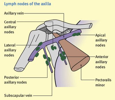
Image 1: Fordyce spots on upper lip

Picture Source : hkma.org

How are they diagnosed?

Most diagnosis efforts will be to rule out sexually transmitted disease as well as cancerous growths. Biopsy and palpitation may be done to check that it is none of that diagnosis. (4)

What causes Fordyce Spots?

  • Technically they are thought to be caused by sebum (natural oils of the skin)
  • They are naturally occurring and present in most adults.
  • Sometimes they are not even seen unless the skin is stretched.
  • They are also more noticeable with those who have greasy skin type.(4)
  • They occur more as one age and are noticed most in elderly people. (5)

How can you prevent Fordyce Spots?

They are not preventable as they are naturally occurring (1,3,4) A recent study has shown that they may be caused by high fat intake in the diet. It is possible that a low-fat diet could prevent and decrease force spots (8)

Where are Fordyce Spots located (Pictures) ?

More specifically Fordyce spots can show at times on the penile shaft, the scrotum, and even the labia for females. As stated before these are natural and appear on many adults.

Fordyce spots located on or around the mouth.photoPicture 2 : Fordyce spots located on or around the lips (mouth)
Image Source : www.dermnetnz.org

Fordyce spots on the penile shaft picture 
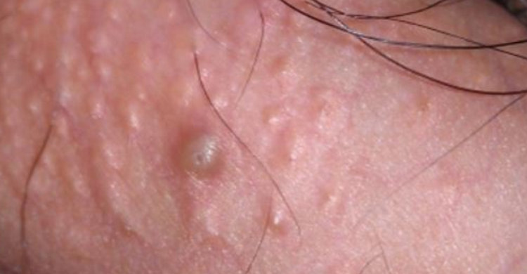
In this image, you can see the difference between natural Fordyce spots on the penile shaft as well as one that was irritated by the patient “picking” at the spot.

Fordyce spots on the penile shaft image photoThis image also shows the natural appearance of Fordyce spots on the penile shaft.

Fordyce spots show up on the labia of the female

This image above shows how the Fordyce spots show up on the labia of the female.

Do Fordyce Spots go away?

They could go away with the treatments noted above but will last anywhere between weeks to months.  In all cases, it is a good idea to get them looked at by a doctor to rule out any sexually transmitted disease. (3)

Are Fordyce Spots contagious?

They are often confused for genital warts which are contagious but Fordyce spots are not contagious and are not sexually transmitted. (3,5,7) nAlthough they are not contagious they are as common as to be in 80-90% of adults.  They are often not noticed until puberty due to changes in skin color during hormone changes. (3)

Are Fordyce Spots itchy?

No, the spots should not be itchy and in the case that they are medical attention is needed.

How to get rid of fordyce spots (Treatment and Remedies)

In reality treatment for Fordyce spots is not necessary because it is not an infection and is normal for many adults. Some medical professionals may even suggest that you do not treat them at all. Some treatments may cause more prominent scarring than the spots themselves.  But if you do want to find a way to get rid of Fordyce spots you could investigate the following treatments.

  • Electrodesiccation- this is the same treatment used for skin tags and other harmless skin conditions. It is the use of CO2 to “burn” off the lesions.
  • Pulsed dye lasers- This is usually used for a condition in which the sebaceous glands are enlarged. It is known to be expensive but may decrease the appearance of the Fordyce
  • Micro-punch technique- Is basically a biopsy method used to remove the spots. It is noted to have a high satisfactory result. (1)
  • Some people will try to express the white thick discharge by squeezing the lesion like a pimple. This usually only gets rid of it for a short time and could cause infection. (4,5)
  • Chemical peels such as trichloroacetic acid- Although this may not remove the bumps it can improve their appearance.
  • There are also many home remedies such as:
    • Garlic being consumed as a regular part of the diet
    • Good personal hygiene may help the appearance of the spots.
    • A diet high in Vitamin B complex, A, C, E, D, and K could help to reduce the spots and even help them to disappear. (5)
    • Adding healthy oils to the diet such as fish oil (6)
  • Topical creams may be used such as Tretinoin and acne washes using benzyl peroxide or salicylic acid.
  • There is a homeopathic cream called Anti-Sebum that may also help with the appearance.

Resources:

  1. http://www.medicalnewstoday.com/articles/256918.php
  2. http://medical-dictionary.thefreedictionary.com/punch+biopsy
  3. http://www.emedicinehealth.com/image-gallery/fordyces_condition_picture/images.htm
  4. https://en.wikipedia.org/wiki/Fordyce_spots
  5. http://byebyedoctor.com/fordyce-spots/
  6. http://www.soc.ucsb.edu/sexinfo/article/fordyce-spots
  7. http://www.aocd.org/?page=FordyceSpots
  8. http://mangoboss.com/FordyceSpots-CausesandTop10NaturalRemedies.html


Ischial Tuberosity

$
0
0

Ischial tuberosity has two parts. The ischial comes from the word ischium which is a pelvic bone. On the other hand, tuberosity is a Latin word, which means lump.

A person with ischial tuberosity has a bony protrusion, which carries the weight of the body when sitting. Hence, it is tagged as the sitting bone. Its other names include sitz bone, tuber ischiadicum, and tuberosity of the ischium. When standing, the ischial tuberosity is covered by the gluteus maximus. (1)

A clear image of the ischial tuberosity, pelvic bone, and the legs.image

Picture 1 : A clear image of the ischial tuberosity, pelvic bone, and the legs
Photo Source : classconnection.s3.amazonaws.com

Recommended exercises for patients with ischial tuberosity problems.photo

Image 2 : Recommended exercises for patients with ischial tuberosity problems
Picture Source : exercisesforinjuries.com

Where is the Ischium located in the human body?

Ischial tuberosity is a part of the ramus, one of the primary bones of the pelvic girdle. It is divided into upper and lower regions. It forms the lower and back part of the hip bone. It is located below the ilium and right behind the pubis.

It has three parts:

  1. the superior ramus
  2. inferior ramus
  3. body. (2)

What muscles are attached to the Ischium and what are the functions of the Ischial Tuberosity?

It is a site of attachment for various muscles and ligaments. They are the following:

  • Coccygeus muscles – It starts at the ischial spine and insert at the bottom part of the coccyx and sacrum. The function of this muscle is to support the organ in the pelvis. (3, 4)
  • Levator ani muscle – It is attached to the pubic bones, ischial spines, and levator ani (tendinous arch). Its function is to support the pelvic organs and resist abdominal pressure, which is useful in maintaining urinary and fecal incontinence.
  • Adductor magnus – A large muscle in the thigh area that is situated right next to the hamstrings. It is the one responsible for the lateral and internal rotation of the upper leg.
  • Biceps femoris, semimembranosus, and semitendinosus are also attached to the ischium. It extends the hind limb when kicking or propelling. (5, 6)
  • Semimembranos muscle – The middle of the three hamstring muscles in the back of the thigh which aids in the extension of the hip joint and bend the knee joint.
  • Sacrotuberous ligament – It anchors the sacrum to the hip bones to prevent tilting.

What is Ischial Spine and its purpose?

The ischial spine is a bony projection into the pelvic outlet from the ischial bones forming – lower border of pelvis. It gives attachment to the sacrospinous ligament and coccygeus muscle. The pudendal nerve goes via dorsal side to ischial spine. It can be palpated through the rectum or vagina. It is the point for needle tip while giving the pudendal nerve block.

Ischial Tuberosity Pain

Ischial tuberosity supports the weight of the body during sitting position. It adjoins different muscles, especially those have to do with the legs.

Athletes commonly used ischial tuberosity, especially for jumping, cycling, running, skating, and soccer. These activities can cause train on the muscles on the legs leading to ischial tuberosity pain. The pain can be very severe.

It will cause the muscles to pull which results to inflammation and injury. Extended sitting could lead to worsening of the condition. Severe cases could lead to detachment of the muscles and even tearing of the ligaments.

What causes Ischial Tuberosity pain?

Extended overload can lead to ischial tuberosity pain. Athletes experienced pain because of the continuous exaggeration of the leg muscles including the hamstring and adductor muscles of the thighs. The pain can be felt in the lower part of the buttocks. Severe pain can be felt even when running and walking fast. Prominent pain could lead to swelling. Pain in any form could mean injuries to the following:

  • Tear in the hamstring muscles
  • Ischial tuberosity tendonitis at the hamstring muscle
  • Stress fracture to the ischial tuberosity
  • A bruise at the hamstring attachment

Sitting on hard surfaces for a long period of time can cause damage to the ischial tuberosity. It could lead to ischial bursitis. The bursa is a fluid-filled sac located in the tuberosity. It acts as a cushion between the pelvis and the tendons nearby to prevent damage.

Sitting for extended period of time could lead to inflammation of the bursa as characterized by pain and tenderness in the hips and buttocks area. It could possibly lead to sciatica wherein there is numbness and pain, which could spread into the legs. (3, 4)

Treatment

Injuries to the ischial tuberosity can be very painful. Hence, the patient is advised to rest and allow the area to heal. Pain is managed using pain medication. Cold compress is used to significantly reduce the swelling and inflammation.

Physical therapy is also helpful to get the muscles back to its pre-injured state. It also strengthens the muscles. For patients suffering from ischial bursitis, they are advised to sit on the padded surface. For severe damage, a corticosteroid injection is needed. Surgery can also be done when deemed necessary. (1, 2)

Other treatment management methods

  • Exercise – Free hand exercise or mild physical exercise helps in pain management. However, you should avoid doing heavy exercises as it could lead to severe pain.
  • Physiotherapy – It helps but does not provide permanent solution to the problem. However, it helps in providing temporary relief from the pain.
  • Medicines – Various medications are prescribed for the treatment and management of ischial tuberosity. Analgesics are used to reduce the pain. However, analgesics are for short term course as prolonged use has adverse effects to the body.
  • Prolotherapy – A sugar solution is injected to the damaged tissue to create a fluid flow onto the affected tissues. It increases the flow of blood and repairs the damaged part by boosting the function of the immune system on the affected area of the body. This procedure allows the growth of new collagen thereby helping in the restoration of the injured ligaments in the joints.
  • Stem Cell Therapy – This procedure produces inflammation to the affected ligaments thereby promoting self-restoration of the damaged tissues and to significantly reduce the pain. (3, 4, 5)

Ischial Tuberosity Exercises

  1. Leg Curls – Performing leg curls with the knees turned outwards can strengthen the hamstrings. You can curl the legs using an outdoor bike or an exercise cycle. There is a leg curl machine specially made to target the hamstrings.
  2. Adductor Machine – Adductor starts on the ischial tuberosity specifically in the adductor magnus muscle. Adductor machine strengthens the adductors.
  3. Hamstring Stretch – Tight adductors and hamstrings can pull the ischial tuberosity causing pain and injury. To relieve the tension, the muscles should be stretched and the flexibility of the hip should be increased.
  4. Frog Adductor Stretch – Adductor stretch helps relieve strain on the ischial tuberosity. Loose adductors can be tightened using stretching exercises. Frog adductor stretch improves the flexibility of the groin and inner thigh area. (6, 7, 8)

References:

  1. http://www.knowyourbody.net
  2. https://en.wikipedia.org
  3. ehealthhall.com
  4. www.boundless.com
  5. www.wisegeek.org
  6. www.innovateus.net
  7. www.livestrong.com
  8. www.caringmedical.com

Rectus Abdominis Muscle and Exercises

$
0
0

Rectus abdominis is popularly known as the abs muscle. It is a pair of long and flat muscle extending vertically along the entire length of the abdomen just near the umbilicus.

The muscles have string of four fleshy muscular bodies that are joined by narrow bands of tendon. It gives the muscles a lumpy appearance when tensed and defined. It is the reason why the rectus abdominis muscle is called six-pack abs. Rectus abdominis are Latin words which means straight and abdominal. (1)

Rectus Abdominis Anatomy

Rectus abdominis muscle extends inferiorly from the pubic crest, pubic symphysis, and pubic tubercle. It surpasses the metastenum. The muscle inserts into the fifth, sixth, and seventh ribs of the costal cartilages by three different parts.

Origin and Insertion

rectus abdominis origin and insertion

Tent of the muscles, especially during contractions. Three bands of connective tissues crossed the rectus abdominis. It is called the tendinous intersections, which is situated just at the umbilicus level. he rectus sheath encloses the flat muscles and hold the muscle in place. It does not restrict the movem

Rectus abdominis is a Mathes and Nahai Type 3 muscle, which has two dominant pedicles. It has a rich supply of arterial blood vessels and consists of 53.9% type 2 fibers. The upper portion of the muscle contains blood from the superior epigastric artery. (3)

rectus abdominis muscle.picture

Image 1 : Rectus abdominis muscle
Photo Source : www.mindandmuscle.net

Muscle Anatomy video

crunching exercises to significantly improve the appearance and strength of the rectus abdominis muscle.image
Picture 2 : Crunching exercises to significantly improve the appearance and strength of the rectus abdominis muscle
Image Source : media-cache-ak0.pinimg.com


A woman performing rectus abdominis muscle exercises using a stability ball.photo

Figure 3 : A woman performing rectus abdominis muscle exercises using a stability ball.
Picture Source : positivemed.com

What is the function and action of the Rectus Abdominis Muscle?

Rectus abdominis muscle helps in various bodily functions. Some of the notable functions of the rectus abdominis muscle include the following:

  • It helps human in maintaining proper posture.
  • It helps in activities that involve flexing of the lumbar region of the vertebral column such as performing abdominal exercises like crunches. (2)
  • It aids in breathing and respiration, especially when it comes to forced exhalation after performing an exercise.
  • It can stretch the abdominal wall and at the same time compresses the abdominal contents. The compression of the abdominal cavity is useful in emergency situations such as the Valsalva maneuver. (4)
  • It helps stabilize the pelvis to allow the lower limb muscle to work properly.
  • It controls the tilt of the pelvis during contraction. (3)
  • It helps in flexing the spine to the front down and forward towards.
  • It helps in the flexion of the thoracic spine and lumbar spine.

What is the Rectus sheath?

The rectus sheath contains the rectus abdominis and the pyramidalis muscles. It is the fibrous condensation of the aponeurotic layers (shiny white tendons) on the anterior portion of the abdominal wall. Aside from the rectus abdominis, the sheath also contains the epigastric vessels, subcostal and intercostal vessels and nerves. (7)

Injury involving the Rectus Abdominis Muscles

The rectus abdominis muscle is prone to injury or damage, especially during extreme workout. Athletes usually experience muscle strain of the abdomen. The muscles of the abdomen can be ruptured or torn depending on the severity of the injury. There will be a notable pain and discomfort in the abdominal area because of the injury.

There are instances when a person will suffer from aggressive fibromatosis or also known as desmoids tumors in the right rectus muscle. The tumor can be detected through a CT scan or MRI scan image. A person may also suffer from a condition called Diastasis Rectus Abdominis (DRA), which causes the rectus abdominis muscles to separate into half. Common symptoms for DRA include epigastric hernia. Some medical reports showed a possibility of endometriosis of the rectus muscle.  (3)

Rectus abdominis strain is one of the common conditions and caused by overstretching of the muscle. People who perform strenuous exercises have the tendency to experience rectus abdominis strain. To prevent muscle strain, you should warm up prior to exercising.

Do not forget to cool down afterwards. You should avoid sudden twisting motions as well as lifting too heavy objects. Rectus abdominis injury is common to swimmers, weight lifters, and those playing hockey and skating. It is also common to overweight people. (8)

Levels of injury to the Rectus Abdominis Muscles

  • First-degree strain – The rectus abdominis is abnormally stretched but is still intact.
  • Second-degree strain – There is a severe stretching of the muscle that resulted to a partial muscle tear.
  • Third-degree strain – There is a complete tear of the rectus abdominis muscle to the point that the muscle separates from the tendons. There is a possibility that the patient will develop a hernia (an abnormal protrusion of the intestines and connective tissues through the rectus abdominis and other muscles in the abdomen.

What are the symptoms of Rectus Abdominis strain?

  • Pain in the abdominal muscle area after performing vigorous activities like running, striking a ball, and prolonged sitting.
  • Pain while coughing, laughing, or sneezing.

Recommended Rectus Abdominis workout : Exercises

There are exercises you can do to significantly improve the appearance and functions of the rectus abdominis muscles. These are the following:

  • Sit-ups – This exercise aims to strengthen the rectus abdominis muscle. It is a simple exercise that is beneficial not just to the rectus abdominis but to other muscles of the body. Lye on your back with your knees bent. Your hand should be behind your head as you are carefully lifting your shoulder blades off the floor. You won’t need any exercising tools or equipment to perform sit ups. The principle is to basically rely to the weight of your body.
  • Stability ball – This exercise provides unstable surface for the rectus abdominis training. Using the stability ball, you can perform a variation of exercises such as sit-ups while on the ball. It enables you to lower your upper body beyond a parallel position to significantly increase the movement during abdominal training. Using the stability ball, lie face down on the ball and place your hands on the floor with your arms straight. Put your hands away from the ball until the ball sits against your shins. Tuck by bending the knees and keep the gently roll the ball towards your chest.
  • Static/Non-movement contractions – It strengthens the rectus abdominis. You can perform this exercise while sitting or even lying down. Just tighten your stomach by pulling the navel towards the spine.
  • Planking – This exercise helps in strengthening the core. Planking for a few seconds daily will make a huge difference in your rectus abdominis muscle. (8)


References:

  1. www.innerbody.com
  2. www.reference.com
  3. http://www.patienthelp.org
  4. http://www.shapesense.com
  5. http://thewellnessdigest.com
  6. www.betterhealth.vic.gov.a
  7. www.kenhub.com
  8. http://www.livestrong.com
Viewing all 48 articles
Browse latest View live