Quantcast
Channel: Dr. Jackie Te RN, MD – Health Fixit
Viewing all 48 articles
Browse latest View live

Growth Factor

$
0
0

What is a growth factor? Definition

It is a naturally occurring substance that simulate the growth of cells. It also promotes healing, proliferation, and cellular differentiation.

It acts as a signalling molecule between the cells. Growth factor is a subset of cytokines. It can be secreted by neighboring cells as well as distant glands and tissues. (3)

How growth factor stimulates growth?

Growth factors stimulate growth through the endocrine, autocrine, and paracrine mechanisms. Growth factors bind their receptors on the target cells’ surface.

However, there are a few factors that could affect the growth of target cells such as the type of receptors, transduction of intracellular signal, number of target cells, the binding ability of growth factor to extracellular matrices, growth factor concentration, and extracellular matrices degradation. (2, 4)

A pathophysiologic presentation of growth factors.image

Photo 1: A pathophysiologic presentation of growth factors.
Picture Source: www.hgh-pro.com

Growth factor serum, one of the popular growth factor supplements today.photo

Image 2: Growth factor serum, one of the popular growth factor supplements today.
Picture Source: www.dermacaredirect.co.uk

What are growth factors types?

Insulin-like growth factor 1 (somatomedins)

It stimulates cellular growth by mediating the secretion of growth hormone from the pituitary gland. It is abundant in the body following birth and play a very important role in the formation of proteins.

Epidermal growth factor (EGF)

it promotes growth of the epithelial cells.

Platelet derived growth factor (PDGF)

It stimulates the growth of muscle and connective tissue cells.

Vascular endothelial growth factor (VEGF)

Stimulates the blood vessel formation

Nerve growth factor (NGF)

It stimulates the growth of neuronal cells

Erythropoietin

It stimulates the growth of red blood cells. It also plays a very important role in the treatment of anemia secondary to chronic kidney failure.

Granulocyte colony stimulating factor (GCSF) and granulocyte macrophage colony stimulating factor (GMCSF)

They stimulate the production of white blood cells in patients with cancer.

Fibroblast growth factor (FGF)

They are primarily involved in embryonic development, wound healing, angiogenesis, and endocrine signalling pathways.

Growth Factor Classes

  • Class I – These are growth factors that interact with a particular type of receptor at the surface of the cell. Examples of class I growth factors are platelet derived growth factor (PDGF), epidermal growth factor (EGF), and growth hormone (somatotropin).
  • Class II – They are cell surface hormone receptors.
  • Class III – They are a large group of intracellular signal transmitters.
  • Class IV – They are proteins that bind to the specific DNA segment or also called nuclear transcription factors. (3, 5, 6)

The effects of growth factor in the body

  1. Growth factors are proliferation regulators. Cells need growth factor to proliferate. Only a few cells can make it through without the growth factor.
  2. Growth factors regulate differentiation (both positive and negative). The actions of some type of growth factors have a lot to do with the development of the cells.
  3. They regulate cell migration and path finding.
  4. They also regulate the growth, survival, and death of cells. (4, 5)

Growth factor supplement

The body’s growth factor level can be affected by various bodily and health conditions. To supplement the body’s need for growth factor, various growth factor supplements were created.

It is important to supplement the body’s need for growth factor because it does not only stimulate cellular growth, but aids in various functions of the body. It plays a major role in cellular differentiation, hastens the healing process, and cellular proliferation, to name a few. (2)

There are many growth factor supplements today. The most popular ones include the following:

Human growth factor serum

It is a powerful nutritional supplement. Many bodybuilders and athletes take it because it can significantly enhance their physical performance, increase muscle mass, and improve their endurance and stamina.

As a matter of fact, the growth factor serum is one of the vital components of face cream. It also helps counteract the aging process.

How is it possible? Growth factor serum has a high level of epidermal growth factor. When you massage it in the skin, it will create a silky sensation. It expedites tissue regrowth and balances the moisture of the skin.

It also protects the skin from free radicals. It nourishes the skin and provides protection from cellular degradation. Growth factor serum is also one of the primary components of peeling treatments. (2, 3, 9)

Chlorella growth factor (CGF)

It is one of the most popular growth factor supplements today. It is derived from chlorella algae, which is considered a super food. It has anti-ageing, immune boosting, and detoxifying property. It hastens cellular repair. It makes you grow fast.

The growth ability of chlorella growth factor is what makes it one of the preferred supplements today. Kids grow faster and they become healthier when taking chlorella growth factor supplement on a day to day basis.

Take a look at the benefits of chlorella growth factor supplement:

  • Chlorella growth factor plays a very important role in detoxification. It helps flush out harmful toxins from the body.
  • It promotes cellular healing and repair. Damaged tissues will be able to bring back to its pre-damaged state.
  • Chlorella growth factor increases growth spurts. A daily intake of chlorella growth factor promotes rapid growth in children.
  • It counteracts the effect of aging. It improves the condition of the hair, nails, and skin.
  • It boosts the body’s ability to fight diseases. It stimulated the production of interferons, a type of immune protein the body produced to combat pathogens.
  • Chlorella growth factor improves the number of healthy bacteria in the gut. A regular intake of chlorella growth factor can help improve the digestive processes of the body.
  • Chlorella growth factor boosts the body’s energy level. It improves the body’s metabolism by stimulating the production of enzymes, proteins, and energy. (6, 7, 8)

Growth factor as the primary component of skin creams

Growth factors are proteins that help regulate the growth, differentiation, and proliferation of cells under a controlled condition. They play a crucial role in maintaining a healthy skin structure. All cell types that make up the skin layers secrete growth factors including the epidermis and dermis.

Growth factors in the skin stimulate biochemical pathways to promote repair and regeneration of skin tissues. They facilitate collagen and elastic fiber formation, which makes the skin supple and soft. Skin care products containing growth factors reduce the formation of fine lines and wrinkles.

They even out pigmentation and age spots. They keep the skin smooth and tight. (1, 2, 10)

References:

  1. https://en.wikipedia.org
  2. www.sinobiological.com
  3. https://themedicalbiochemistrypage.org
  4. www.getgrowthfactor.com
  5. www.sciencedirect.com
  6. www.abmgood.com
  7. www.skinmedica.com
  8. www.growthfactor.com
  9. www.raybiotech.com
  10. Epidermal Growth Factor: Methods and Protocols edited by Tarun B. Patel, Paul J. Bertics

Medial Knee Pain

$
0
0

The knee is a large and complex joint. It connects the femur to the tibia. It also has other surrounding structures like the tibia and kneecap.

The knee plays a very important role in various bodily functions and movements such as walking, running, and jumping. The complexity of the structure of the knee makes it susceptible to pain. The middle part of the knee experiences most of the pain.

Medial knee pain occurs for a variety of reasons. The common medial knee pain causes include the following:

MCL Tear (Medial Collateral Ligament)

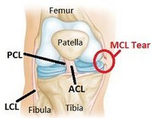
some or all of the fibers of the MCL on the inner side of the knee are damaged. The patient complains of a sudden yet severe medial knee pain. There is also bruising, swelling, stiffness, and instability. The pain is worsened when walking, climbing the stairs, or bending the legs. (1, 2)

Meniscus Tear

It is also known as the medial cartilage tear. It is characterized by a tear in the cartilage lining of the inner side of the meniscus (knee joint). The pain is sudden. The patient finds it difficult to straighten the legs. There is also swelling and instability. Simple activities can trigger medial knee pain such as walking, climbing the stairs, squatting, and running. (3)

Pes Anserine Bursitis

It is the less common cause of medial knee pain. It is characterized by inflammation of the pes anserine burse, a small fluid-filled sac. If there is an inflammation, the fluid that reduces friction in the knee bones gets dry leading to pain and inflammation, especially when moving.

The onset of the pain is sudden and gradually intensifies. The pain is more intense when the patient tries to climb the stairs. It commonly affects overweight middle-aged women as well as swimmers and runners.

Arthritis

It is a medical condition characterized by wear and tear of the cartilage that lines the medial side of the joints. The one that causes medial knee pain is osteoarthritis. Medial knee pain worsens as the person grows old.

Patients 50 years old and above are prone to osteoarthritis. The clinical manifestations include inside knee pain, a stiffness of the joints in the early morning, reduced movements of the legs, and swelling. The patient also complains of chronic medial knee pain. The patient is aggravated by cold weather and prolonged activity or rest. The severity of symptoms depends on whether osteoarthritis is mild, moderate, or severe. (4, 5)

Medial cartilage meniscus injury

A torn meniscus causes pain on the inside structure of the knee. This type of injury is common in athletes, especially in contact sports such as basketball.

Knee contusion

It occurs because of a sudden impact on the side of the knee. In layman’s term, it is called bruising of the knee. It may sound simple on the outside but it could cause damage to the soft tissues and even to the bones. (2)

Medial knee pain is common because the muscles in the knee area are extremely sensitive. If the muscles become weak or tight, it affects the way the knee moves. More force will go through the inner side of the joints leading to damage to the inner side of the knee.

Anatomy of the knee joints pictures Image 1: Anatomy of the knee joints.
Photo Source: progressivephysio.com

MCL tear which is one of the cause of medial knee pain photo

Picture 2: MCL tear which is one of the cause of medial knee pain.
Image Source:  www.knee-pain-explained.com

A knee brace, supports the knee, keeps the knee in placed, and prevent further injury to the knees image

Photo 3: A knee brace, supports the knee, keeps the knee in placed, and prevent further injury to the knees.
Picture Source: www.physioroom.com

Medial knee pain treatment

Inflammation is the body’s way to cope with injury. When it comes to treating medial knee pain and knee pain in general, one of the primary goals is to break the inflammatory response. If the inflammation is not resolved the soonest time possible, it could lead to chronic medial knee pain and severe injury to the knee.

It makes the treatment quite difficult considering that the infection has already advanced. The goal of the medial knee pain treatment is to control the substance that causes inflammation and limiting movements that could further injure the knee. (3, 4, 5, 6)

The first treatment approach for knee pain is protection, rest, ice, compression, and elevation (PRICE).

  • Protection – It is important to protect the knee from further trauma. To protect the knee, a split or knee pad should be used. A pad over the knee can help control the symptoms of knee injuries.
  • Rest – Do not move the injured knee to significantly reduce the strain and give ample time for the injured knee to heal.
  • Ice – Cold compress helps reduce swelling and inflammation.
  • Compression – A medial knee pain brace reduces swelling, keep the alignment of the patella, and keep the joint mechanic intact.
  • Elevate – By simply elevating the knee, swelling can be reduced and blood circulation will be improved significantly. (4, 6, 7)

Pain Management

Over the counter pain medications are used to control the pain. If the pain does not go away, then the doctor will prescribe stronger pain medication.

Pain medications do not only control the pain but also help alleviate inflammation and facilitate in the breaking of an inflammatory cycle. If you have stomach ulcers or bleeding problems, you should not use non-steroidal anti-inflammatory drugs.

Medial knee pain exercises

There are various exercises for medial knee pain. However, exercises should only be done if the injury is not painful to touch and not painful to stretch. If there is an intense pain even when the patient is at rest, then the patient should refrain from doing any types of exercise.

Before starting any forms of exercise for the knee pain, you have to make sure you consult your doctor. If you exercise without consulting your doctor, you could be adding injury to the knees. (7, 8, 9, 10)

When should you call out for professional help?

If you have tried all the treatment remedies mentioned above but still the pain does not go away, then it is high time to consult your doctor. The doctor or physical therapist will further evaluate the pain.

The more you need to talk to your doctor if the knee pain gets intense, you have a fever, and you are feeling sick. If there are signs of infection, then you need to go to the emergency room to immediately treat the underlying condition and prevent secondary infection. (2, 6, 10)

References:

  1. http://www.sportsinjuryclinic.net/sport-injuries/knee-pain/medial-knee-pain
  2. https://www.healthline.com/health/inner-knee-pain
  3. http://www.knee-pain-explained.com/medial-knee-pain.html
  4. http://www.londonorthopaedic.com/medial-knee-pain/
  5. https://www.injurytreatment.com.au/injury-information/knee/medial-inside-knee-pain/
  6. https://www.braceability.com/blog/types-of-knee-pain-anterior-posterior-medial-lateral-knee-pain/
  7. https://www.webmd.com/pain-management/guide/knee-pain-overview#1
  8. https://www.thekneepainguru.com/inner-knee-pain/
  9. https://en.wikipedia.org/wiki/Medial_knee_injuries
  10. http://mobilitymastery.com/how-to-relieve-medial-knee-pain-low-back-si-pain-groin-pain-more

Odynophagia

$
0
0

What is Odynophagia? Definition

Odynophagia is a medical term for painful swallowing. It is often confused with the term dysphagia, which means difficulty swallowing. With odynophagia, the patient complains of pain when trying to swallow.

The pain can be felt in the mouth, throat, and/or esophagus. If the patient complains of pain when swallowing, it is an indicator that something is not right in the body. It is important to consult your doctor so that the underlying cause will be determined and the patient will be able to receive the appropriate treatment. (1, 2, 3)

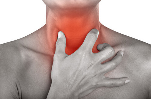
A painful throat is an indicator of a painful swallowing (odynophagia) pictures

Photo 1: A painful throat is an indicator of a painful swallowing (odynophagia).
Image Source: diseasespictures.com

A tumor in the esophagus, which is one of the reasons for painful swallowing image

Picture 2: A tumor in the esophagus, which is one of the reasons for painful swallowing.
Photo Source: yoursurgery.com

Odynophagia Causes

  • Cancer – When a patient complains of chronic painful swallowing, the doctor will rule out esophageal cancer. The tumor develops in the esophagus. There are various reasons why tumor develops in the esophagus such as alcohol abuse, chronic smoking, and persistent heartburn. In rare instances, esophageal cancer can be hereditary.
  • Gastroesophageal reflux disease (GERD) – the lower sphincter in the esophagus is not closing properly. This causes the stomach acid to leak back into the esophagus. One of the symptoms of GERD is painful swallowing, chest pain, and heartburn. (3)
  • Candida infection – This fungal infection can affect the mouth. It will spread and affect the esophagus. That’s why patients with candida infection complain of painful swallowing.
  • Ulcers – They are sores that can affect the oral cavity particularly the mouth, throat, esophagus, and stomach. Factors that can increase the risk of ulcers are a chronic use of anti-inflammatory drugs and untreated Gastroesophageal reflux disease.
  • HIV – People with HIV are immunocompromised. They are prone to infection. The retroviral drugs used to treat HIV can increase the possibility of painful swallowing. (4)
  • Side effects of treatment – Some forms of treatment can cause painful swallowing such as radiation therapy for cancer and side effects of prescription medications.
  • Object stuck in the throat – Small sharp objects stuck in the throat can significantly affect your ability to swallow. It causes painful swallowing. (4, 5)

Difference between dysphagia and odynophagia

These two medical terms are somewhat related that’s why a lot of people used it interchangeably. However, odynophagia is different to that of dysphagia.

Dysphagia is when a person experiences difficulty in swallowing. The swallowing difficulty can occur on a regular basis and is common in older people. Not all people with swallowing difficulties experienced pain.

If you have difficulty swallowing but don’t have any pain at all, then the condition is called dysphagia. On the other hand, odynophagia is the term used to describe a painful swallowing. (5, 6, 7)

Odynophagia Symptoms

  • Pain when swallowing
  • Shortness of breath
  • Pain in the oral cavity that intensifies when a person tries to swallow
  • The patient complains of globus sensation. What is the globus sensation? It is the feeling of having a lump or obstruction in the throat when there is really none. (7, 8)

Diagnosis

Endoscopy is the commonly used procedure to diagnose odynophagia. A small lighted camera (endoscope) is placed in the throat so that the doctor can visualize the esophagus. The doctor will also ask you to swallow during the procedure.

Other tests will be ordered to accurately diagnose the underlying cause of odynophagia.

Odynophagia Treatment

Odynophagia treatment guidelines include the following:

  • Drug therapy – depending on the underlying cause, the doctor will prescribe medications such as medications for GERD to prevent stomach acid to go back into the pharynx and esophagus. The medicines are used to target the underlying cause of odynophagia. If it is caused by candida infection, then an antifungal agent should be used.
  • Surgical approach – if the cause of painful swallowing is a tumor, the doctor will perform a surgical procedure to get rid of the tumor cells. If you have GERD and the condition does not improve despite aggressive drug therapy, then the doctor will perform surgery for GERD.
  • Time – If there is no underlying cause for odynophagia, then maybe you should give it some time to resolve on its own. Painful swallowing is common, especially after severe allergies or cold. When the infection completely goes away, your swallowing will improve significantly. (4, 9, 10)

Outlook

Many underlying health conditions for odynophagia will significantly improve if detected and treated early. If you are experiencing swallowing difficulty, then you should inform your doctor the soonest time possible.

If the condition is left untreated, it could lead to more serious complications. If you have a painful swallowing, your eating habit will be affected. You will eventually lose weight. The body will not receive the nourishment it needs. It could lead to dehydration, malnutrition, and anemia. It could also make you susceptible to different types of illnesses. (3, 4)

References:

  1. https://en.wikipedia.org/wiki/Odynophagia
  2. https://www.healthline.com/health/odynophagia
  3. http://www.healthhype.com/swallowing-pain-causes-of-odynophagia-painful-swallowing.html
  4. http://www.clinicaladvisor.com/hospital-medicine/dysphagia-and-odynophagia/article/600672/
  5. https://www.doctorshealthpress.com/health-articles/odynophagia-painful-swallowing/
  6. http://www.ehealthstar.com/painful-swallowing-odynophagia.php
  7. https://www.medicalopedia.org/911/dysphagia-vs-odynophagia/
  8. https://heydoctor.org/odynophagia.html
  9. https://www.singhealth.com.sg/PatientCare/ConditionsAndTreatments/Pages/odynophagia-pain-on-swallowing.aspx
  10. http://www.medicotips.com/2013/04/why-painful-swallowing-odynophagia.html

What side is your appendix on

$
0
0

Appendicitis is the inflammation of the appendix. What is an appendix? It is a tiny sac attached in the cecum (first section of the colon) in the right lower quadrant of the abdomen.

It is a vermiform appendix because of its resemblance to a worm. It is anatomically positioned in the right iliac region of the abdomen. (1, 2)

what side is your appendix on location picture image photo

Image 1: The anatomical presentation of the appendix. It is an appendage, which resembles like a worm.
Photo Source: images.onhealth.com

Other names for appendix

  • Vermiform appendix
  • Cecal appendix
  • Vermix

The functions of the appendix

  • It contains lymphatic tissue, which plays a very important role in immune function.
  • It functions as a storage area for good bacteria.
  • It hastens the recovery from illness by allowing the good bacteria to re-colonize the intestines. (1, 2, 3)

what side of the body is your appendix appendicitis pain location image picture photo

Picture 2: The image shows the inflammation of the appendix or medically known as appendicitis.
Image Source: cdn.universityhealthnews.com

The progression of appendicitis throughout the course of time appendix location image photo picture

Photo 3: The progression of appendicitis throughout the course of time.
Picture Source: edition.channel5belize.com

Where is the appendix located?

What side of your body is your appendix? It lies at the junction of the small and large intestines. Ti is a finger-like pouch located in the right lower quadrant. It is four inches long and as thick as that of a pencil. (2, 4)

appendix location with mcburney point anatomy picture

Picture 4 : Appendix location with Mcburney’s point

What side your appendix on? It is found on the right side of the body, on the right hand side of the abdomen, just above the waist. To find out the exact location of the appendix, you should do these steps:

Locating the appendix using the hip bone

  • Palpate and place your thumb on the top part of your right hand hip bone.
  • Place the pinky finger on the side of the pubic bone.
  • Extend your index finger and there you will locate the exact position of the appendix.

Locating the appendix using the McBurney’s point

The location of the appendix is called the McBurney’s point. To locate the McBurney’s point, create an imaginary line from the navel to the right side of the hip bone. The location of the appendix is about 2/3 away from the navel.

The exact location of the appendix is helpful in accurately diagnosing appendicitis. The doctor applies pressure to the McBurney’s point to confirm if the abdominal pain is caused by an inflamed appendix. (1 4, 6)

What side does your appendix hurt on?

Pain in the abdomen is one of the clinical manifestations of appendicitis. However, there are other health conditions causing abdominal pain. Hence, it is important to know which side of the body is inflamed and in pain if you have appendicitis.

  • Appendicitis related pain usually begins at the center of the belly.
  • As the condition progresses, the entire abdominal wall is swollen and irritated leading to a localized pain at the right lower quadrant of the abdomen.
  • A rebound tenderness is used to detect the location of the appendix pain. The doctor will gently press the abdominal area to watch out for signs of tenderness. Once the doctor releases the pressure, the pain in the abdominal area gets worst.
  • The location of appendicitis pain can also be pinpointed when the patient takes a deep breath or simply coughs or sneezes. (2, 5, 8)

What causes appendicitis?

The exact cause of appendicitis is unknown. However, many health experts believed that it has something to do with the blockage in the entrance of the appendix. It could be a small piece of fecal matter or a foreign substance that blocks the appendix.

If the blockage is not removed, it will cause pressure to the appendix. The appendix will swell and will eventually burst. (2, 3, 4)

Clinical manifestations

  • Pain in the abdomen that is quite difficult to pinpoint.
  • The patient usually complains of pain the mid portion of the abdomen and radiates to the right lower abdominal quadrant.
  • Loss of appetite
  • Weight loss (5)
  • Nausea and vomiting
  • Fever
  • Unable to pass gas
  • Fatigue (4)

Prevalence rate

Nobody is spared from appendicitis. Anybody can be a target. However, it is most common in people aged 10 years old and 30 years old. Young children and older people are prone to appendicitis complications.

Therefore, it is important to detect the condition the earliest time possible to prevent complications. (4, 5, 6)

Appendicitis in children

Appendicitis in children have varying reasons. Children usually complains of stomachache but it could be associated with other conditions such as gas, constipation, and mild food allergy. An abdominal pain that spreads to the right lower quadrant of the abdomen is one of the cardinal signs of appendicitis.

Other symptoms affirmative of appendicitis in children include abdominal pain that gets worst with movement, coughing, sneezing, nausea, lack of appetite, and deep breathing/labored breathing. Very young children have distinct set of symptoms too such as abdominal pain, fever, loss of appetite, and vomiting (children 2 to 5 years old). (6, 7, 8, 9)


One of the diagnostic procedures used to check the condition of the appendix location image photo picture

Image 5: One of the diagnostic procedures used to check the condition of the appendix.
Photo Source: www.acep.org

How to diagnose appendicitis?

To accurately diagnose appendicitis, the doctor will thoroughly examine the patient’s clinical manifestations. The doctor will check the abdomen to detect the exact location of pain. When the doctor pushes down the right lower quadrant of the abdomen, the patient usually complains of moderate to severe pain.

When the doctor removes the pressure on the abdomen, the pain intensifies. To check for the presence of infection, the doctor will order a blood test. An increase in white blood cells indicates that there is an ongoing infection. Additional procedures might be needed such as x-ray and CT scan of the pelvis and abdomen. (7, 8, 9, 10)

Treatment options for appendicitis

An inflamed appendix should be surgically removed. If it burst inside the body, it could cause serious complications. It would put the patient in a life-threatening situation. To remove the appendix, an appendectomy is performed.

It is performed under general anesthesia using the open technique or the keyhole method. (2, 4)

Antibiotics

The patient should receive antibiotics before and after the surgical removal of the appendix.

A keyhole surgery is one of the less invasive procedures for removing appendicitis appendix location image photo picture

Photo 6: A keyhole surgery is one of the less invasive procedures for removing appendicitis.
Picture Source: www.misurgery.com.au

Appendectomy using the open surgery method appendix location image picture photo

Image 7: Appendectomy using the open surgery method.
Photo Source: i.ytimg.com

A laparoscopic procedure in a patient with perforated appendicitis appendix location image picture photo

Picture 8: A laparoscopic procedure in a patient with perforated appendicitis.
Image Source: dccdn.de/pictures.doccheck.com

Surgery

  • Open surgery – It is the surgical technique used if the appendix has already burst. It is also the technique of choice for patient who has had an open abdominal surgery. As the name suggests, a single huge cut is made in the lower right quadrant of the abdomen.
  • Laparotomy – A laparoscopic procedure is when a surgeon makes a cut along the middle of the abdomen. This is the surgical approach for patients with widespread peritonitis. It is a complication of appendicitis. It occurs when the tissues that line the abdomen (peritoneum) become inflamed.
  • Keyhole surgery – It is the most ideal surgical approach for patients whose appendix hasn’t burst yet. A tiny cut in the belly button is made and a special instrument is inserted, which enables the surgeon to completely view the appendix and the surrounding structures. Using a small surgical tool, the appendix is removed. The incision site will be closed using dissolvable stitches. If the doctor used regular stitches, they should be removed after a week. (1, 6, 8, 10)

Recovery time

The recovery period depends on the patient’s overall health condition and the type of surgical approach used. The keyhole technique has a faster recover period than the open surgery and laparotomy. With keyhole surgery, the recovery time is short.

The patient can go home a few days post-surgery. There are instances when the patient can go home 1 to 2 days after surgery. However, if the patient has a complicated surgery such as in the case of peritonitis, the patient will most likely need to spend the entire week in the hospital post-surgery for thorough monitoring. (2, 3, 6, 7)

What to expect after the surgical procedure?

  • Pain and bruising in the incision site.
  • Pain in the tip of the shoulder that could last for a week (keyhole surgery).
  • Short-term constipation (2, 3)

Post-surgical care

  • You should take pain killer and anti-inflammatory as prescribed by your doctor.
  • Avoid strenuous activities.
  • Wound dressing is a must.

During the recovery period, there is a possibility that the patient will not be able to recover completely. There is always a possibility of post-surgical problems.

It is important to keep an eye for unusual signs such as severe pain and swelling, high-grade fever, unusual discharge coming from the wound, and vomiting. If you notice these signs, you should immediately contact your health care provider. (2, 5, 7, 8)

References:

  1. https://www.webmd.com/digestive-disorders/digestive-diseases-appendicitis#1
  2. https://www.medicinenet.com/appendicitis/article.htm
  3. https://www.nhs.uk/conditions/appendicitis/symptoms/
  4. https://www.nhs.uk/conditions/appendicitis/
  5. https://www.healthline.com/health/appendicitis
  6. https://www.medicalnewstoday.com/articles/158806.php
  7. https://en.wikipedia.org/wiki/Appendicitis
  8. https://www.emedicinehealth.com/appendicitis/article_em.htm
  9. http://m.kidshealth.org/en/parents/appendicitis.html
  10. https://www.prevention.com/health/5-warning-signs-your-appendix-might-burst

How to Fix a Dislocated Toe ?

$
0
0

Toes are tiny body parts that are prone to sprain, dislocation, and breakage. A dislocated toe is when the toe bone moves out of its normal position. A dislocated toe occurs when the ligament that holds the bones in the toe together is torn. A dislocated toe rarely occurs. It only happens when there is a strong blow to the toe. (1, 2, 3)

A dislocated toe characterized by redness, swelling, and a bone out of place image photo picture

Image 1: A dislocated toe characterized by redness, swelling, and a bone out of place.
Picture Source: www.paincare.org

severe case of a dislocated toe image photo picture

Photo 2:  severe case of a dislocated toe.
Picture Source: i.ytimg.com

A patient with a severely dislocated toe picture photo image

Image 3: A patient with a severely dislocated toe.
Picture Source: ncrypted-tbn0.gstatic.com

A patient with multiple dislocated toes image photo picture

Photo 4: A patient with multiple dislocated toes.
Picture Source: 3.bp.blogspot.com

Who is prone to having a dislocated toe?

Those who are involved in various sports activities are prone to having a dislocated toe such as soccer players, rugby players, basketball players, dancers, climbers, hikers, and the likes. (2, 3)

Those who are involved in sport-related activities are prone to injuries like dislocated toe image photo picture

Image 5: Those who are involved in sport-related activities are prone to injuries like dislocated toe.
Picture Source: www.trainingcor.com

How do you know if you have a dislocated toe?

How can you tell if your toe is dislocated? What are the signs and symptoms? A dislocated toe is possible is there is a visible misalignment. Most of the time, it is quite difficult to tell if you have a dislocated toe because its symptoms are similar to that of a sprained or a broken toe. The signs and symptoms of a dislocated toe are as follows:

  • Visible misalignment/toe bone is visibly out of place
  • Intense pain that gets worse when you attempt to move the affected toe
  • The toe cannot bear even the slightest pressure
  • The affected toe is stiff and hard followed by pale and cool skin indicating that the blood vessels of the affected toe are damaged.
  • Swelling and bruising (3, 4, 5)

How to fix a dislocated toe?

What is the best way to fix a dislocated toe? It is something that you can do at home or well it requires a surgical procedure? The treatment approach for a dislocated toe varies depending on the severity of the injury. Ideally, treatment should be given to the patient as soon as possible.

A dislocated toe left untreated can progress to the worst scenario, which requires a more advanced treatment. (6, 7)

How to fix a dislocated toe at home?

A dislocated toe should be treated using the RICE method photo picture image

Photo 6: A dislocated toe should be treated using the RICE method.
Picture Source: www.wikihow.com

A buddy tape is a procedure in which the injured toe is taped to the adjacent toe image photo picture

Image 7: A buddy tape is a procedure in which the injured toe is taped to the adjacent toe.
Picture Source: img.webmd.com

First, you need to be certain that the patient has indeed a dislocated toe. Refer to the signs and symptoms mentioned above. If there is indeed a dislocated toe, then you should do the following:

Gentle tug – Gently pop the dislocated toe joint back to its pre-dislocated state. Make sure that you do the procedure right or else you will end up causing further harm. How to do a gentle tug?

  • Run your fingers over the injured toe in a pincer position. Make sure that the direction of your fingers is away from your body. Gently partway up the foot and feel for yourself the exact location of the dislocated toe.
  • Gently tug the misaligned joint away from the toe (away from the knuckle area) until you hear a snap, which means that the joint is back into its original alignment.
  • If you don’t hear a snap, that’s okay. If you reset it correctly, it should be backed to its original alignment.

RICE method – The dislocated toe should be rested, iced, compressed, and elevated. As much as possible, the intense movement should be avoided to avoid causing further harm to the injured toe. To avoid re-injuring the toe, you should splint it or at least try to buddy-wrap the toe to its adjacent toe. (5, 6, 7, 8)

What to keep in mind?

The treatment for a dislocated toe should be carried out within six hours after the toe dislocation. If the dislocated toe has passed the first six hours and no immediate treatment was given to the patient, then the best thing to do is to see a doctor, especially someone specializing in toe-related injury.

Do not attempt to self-adjust way beyond the first six-hour period or else you could end up causing further harm than good. (8, 9, 10)

How long does it take to recover from a dislocated toe?

It would usually take six weeks for the torn ligament to heal. If the injury is severe, the recovery period would take more than six weeks. However, with treatment and proper management and care, the patient’s recovery will be hastened. (3, 5)

Dislocated toe surgery

A case of an open reduction internal fixation of a floppy toe image photo picture

Photo 8: A case of an open reduction internal fixation of a floppy toe.
Picture Source: faoj.org

A dislocated toe surgery is the last resort. It should only be done if the dislocated toe cannot be positioned properly. The best candidates for dislocated toe surgery are those with a floppy toe. What is a floppy toe? It is a dislocated toe in which the toe is in an awkward position.

It usually occurs after the fracture and the joint becomes unstable, which makes it extremely difficult to align the toe manually. The goal of the surgery is to fix the broken toe in place using wires so that the toe remains in its position while it heals.

The surgical procedure is called open reduction and internal fixation. It is the surgery of choice for dislocated and fractured toe, especially if there are multiple toe fractures. After the surgery, the patient should refrain from doing things that can aggravate the toe.

Depending on the extent of the surgery, it would take six to nine weeks for the patient to completely recover. If the toe has been completely healed, the patient can go back sports and activities without the risk of re-injury. (3, 5, 8, 10)

When should you see a doctor?

  1. Contact your doctor if the dislocated toe has passed the first six-hour period and no immediate care was given to the patient.
  2. The pain is extreme that the patient could no longer bear it.
  3. The patient complains of weakness, tingling sensation, and numbness of the toe. More so, if there is visible discoloration.
  4. The dislocated toe does not get better even after religious home remedies. (4, 8)

References:

  1. http://www.sportsinjuryclinic.net/sport-injuries/foot-heel-pain/dislocated-toe-1
  2. https://myhealth.alberta.ca/Health/aftercareinformation/pages/conditions.aspx?hwid=uh3489
  3. https://study.com/academy/lesson/dislocated-toes-symptoms-treatment-recovery-time.html
  4. http://www.thetoedoctor.com/dislocated-toe/
  5. http://www.tandurust.com/bones-and-joints-disorders/dislocated-toe-how-to-treat-it.html
  6. http://www.clinic-hq.co.uk/article_46_Dislocated+Toe
  7. http://www.paincare.org/dislocated-toe/
  8. http://www.winchesterhospital.org/health-library/article?id=912642
  9. http://footcare.ygoy.com/2011/12/01/dislocated-toe/
  10. https://healingfeet.com/foot-care/out-of-place-all-about-dislocated-toes

Prostate Location

$
0
0

The prostate is a gland anatomically situated in between the bladder and the penis. Its size is similar to that of the walnut. Its shape is similar to that of the rounded cone and the base points toward the bladder.

It is both a muscular and a glandular structure. The prostate has ducts that open into the prostatic area of the urethra. It has three lobes, one in the middle and the other two on both sides. (1, 2, 3)

Prostate gland anatomy

The prostate’s anterior end contains two lateral lobes that are round in shape. When viewed transversally, the prostate looks like orange slices. In the posterior part of the lateral lobe is a much smaller lobe containing fibromuscular tissues, which contract during ejaculation.

Another significant structure if the median lobe containing ejaculatory ducts. On the other hand, the posterior lobe creates a thin layer of tissues. (2, 3, 4)

Where is the prostate located

The prostate is in the middle of the pelvis just about two inches in and toward the belly. It is about two to three inches in the male rectum. The prostate gland size is similar to that of the walnut and chestnut. Its weight is between 20 grams to 30 grams. (4, 5)

PROSTATE LOCATION where is the prostate located

The male reproductive system with emphasis of the prostate gland location image photo picture

Image 1: The male reproductive system with emphasis of the prostate gland.
Picture Source: img.webmd.com

A comparison image between a normal-healthy prostate and prostate cancer image photo picture

Picture 2: A comparison image between a normal/healthy prostate and prostate cancer.
Photo Source: www.mvpchiro.com

A rectal examination to detect an enlarged prostate photo image picture

Photo 3: A rectal examination to detect an enlarged prostate.
Image Sourcce: www.naturalcolonrx.com

A comparison image of a healthy and inflamed prostate photo picture

Image 4: A comparison image of a healthy and inflamed prostate.
Picture Source: www.sweet-cures.com

Tissues in the prostate gland

There are two types of tissues in the prostate gland. These are the fibromuscular tissues and exocrine glandular tissues.

  1. Fibromuscular tissue – It is a combination of the smooth muscle tissues and dense irregular connective tissues. It contains collagen fiber that provides strength to the tissues. The smooth muscle allows the tissue to contract so as to expel the fluid during ejaculation. The fibromuscular tissues are found in the outermost layer of the prostate gland.
  2. Exocrine glandular tissue – It is an epithelial tissue responsible for the secretion of semen. Majority of the prostate consists of exocrine glandular tissue. (3, 4, 5, 6)

What are the functions of the prostate gland?

  • The prostate gland is a part of the male reproduction system. Its function is to help secrete a fluid that is part of the seminal fluid (carrying the sperm). During the ejaculation process, the prostate propels the fluid into the urethra.
  • The prostate produces a prostatic secretion, which is a milky mixture of sugars, specifically glucose and fructose, enzymes, and alkaline chemicals. The sugar provides nourishment to the sperm while the enzymes help break down essential protein in the semen. The alkaline material neutralizes the acidic vaginal environment thereby helping the sperm to make it through during implantation.
  • The prostate gland has an ejaculatory duct that facilitates the release of sperm during ejaculation. The duct opens and the smooth muscle tissues in the prostate contract enabling the semen to freely pass into the urethra.
  • The prostate plays an important role in male’s fertility. It has a prostate-specific antigen (PSA) that enables the sperm to swim into the uterus.
  • Prostate gland does a wonderful job of achieving sexual satisfaction. The pumping action of the prostate gland produces a very good feeling making sex more desirable and pleasurable.
  • The prostate gland is the male’s sexual pleasure spot, an equivalent of G spot in the female. Stimulation of the prostate produces a strong sexual response and intensifies the orgasm.
  • The nerves in the prostate help in achieving and maintaining an erection. Prostate erection nerves facilitate swelling and hardening of the penis.
  • Prostate gland filters and facilitates removal of toxins. It protects the sperm and increases the chances of impregnation.
  • A healthy prostate gland reduces the possibility of urinary infection. It is the reason why men are less susceptible to urinary tract infection than women.
  • Prostate gland helps in regulating the production of the male hormone testosterone. The prostate gland has a 5-alpha-reductase enzyme that converts testosterone into DHT. (5, 6, 7, 8)

Prostate Gland Problems

As men grow older, problems in the prostate gland start to appear. It is common in men 50 years old and above, which is why health care professionals strongly suggest that older men should undergo PSA test every year. Common prostate gland problems include:

Prostatitis

It is the inflammation of the prostate secondary to infection. It is usually treated using antibiotics.

Enlargement of the prostate/benign prostatic hypertrophy

It is the abnormal growth of the prostate gland, which is a common condition in older men. Prostate gland enlargement symptoms include difficulty urinating, urine urgencies, especially at night, dribbling after urinating.

The patient is put on a drug that can help shrink the prostate. If the condition is already in the advanced stage, the best thing to do is to perform surgical procedures.

Prostate cancer

It is one of the common forms of cancer in men. What are the 5 early warning signs of prostate cancer? These are frequent painful urination that may contain blood, erectile dysfunction, inability to control the bladder, lower back pain, and decreased semen during ejaculation. What makes you susceptible to prostate cancer? Men older than 50 years old are prone to prostate cancer.

The prevalence is high in African-American men. If any of your immediate family members have had a prostate cancer, then there is a possibility that you can have it too. Don’t you know that your diet can account for the formation of prostate cancer? A high-fat diet makes you susceptible to prostate cancer.

The treatment approach for prostate cancer varies depending on the extent and severity of the condition. If it is in the early stage, a radiation and chemotherapy are used to kill the cancer cells. The body will be hindered from producing testosterone so as to prevent recurrence. Hence, the patient needs to be put in a hormone therapy. Lastly, a surgical removal of the prostate can be done when deemed necessary, especially if the entire prostate is involved. (6, 8, 9, 10)

What happens when you have your prostate removed?

If the prostate is surgically removed, the patient could have urinary incontinence and erectile dysfunction.

References:

  1. https://www.webmd.com/urinary-incontinence-oab/picture-of-the-prostate#1
  2. https://www.emedicinehealth.com/enlarged_prostate/article_em.htm#what_causes_an_enlarged_prostate
  3. https://www.livescience.com/32751-what-does-the-prostate-gland-do.html
  4. https://en.wikipedia.org/wiki/Prostate
  5. https://www.macmillan.org.uk/information-and-support/prostate-cancer/early-prostate-cancer/understanding-cancer/the-prostate-gland.html
  6. https://prostate.net/articles/10-amazing-functions-of-the-prostate-gland
  7. http://www.kidneyurology.org/Library/Urologic_Health.php/Prostate_Enlargement.php
  8. https://www.betterhealth.vic.gov.au/health/conditionsandtreatments/prostate-gland-and-urinary-problems
  9. https://www.nia.nih.gov/health/prostate-problems
  10. https://universityhealthnews.com/daily/prostate/what-is-the-prostate-gland/

Intercostal Muscle Strain

$
0
0

The intercostal muscle is one of the important muscles in the upper cavity. It plays a huge role in stabilizing the upper body and aid in breathing.

The intercostal muscles have three layers: the external, internal, and innermost layer. Because of some activities and health practices, the layers of the intercostal muscles are strained to cause a variety of health issues. (1, 2, 3)

An anatomy of the intercostal muscles, the muscles found in between the ribs image photo picture

Image 1: An anatomy of the intercostal muscles, the muscles found in between the ribs.
Picture Source: digikalla.info

What are intercostal muscle strain symptoms?

  • Upper back pain that is sharp in nature
  • The pain becomes intense when you try to stretch or twist the muscles or when you cough, sneeze or breathe deeply.
  • Muscle spasm and tension
  • The area between the ribs is tender to the touch.
  • Difficulty of breathing as manifested by a small and shallow sip of air.
  • An obvious swelling in between and around the ribs.
  • The pain gets intense with repetitive movements such as swimming and other forms of strenuous activities. (2, 3, 4, 5)

What causes intercostal muscle strain?

  • Injury to the ribs secondary to sports (contact sports).
  • Direct blow to the ribcage such as in the case of a car accident and fall.
  • Twisting of the muscles while lifting heavy objects such as in the case of weightlifters.
  • An intercostal muscle strain as a result of extreme yoga posture, dance position, playing games that require forceful twists such as tennis and golf. (4, 5, 6)

When is the best time to consult your doctor?

You need to carefully observe the extent of the injury, especially if the pain gets severe as time passes by. If the pain is bothersome, to the point of hindering you from performing your day to day activities, then it is high time to consult your doctor.

Intercostal muscle strain diagnosis

The doctor will conduct a series of test to accurately diagnose the condition of the patient.

  • Physical exam – The doctor will conduct a thorough physical exam to detect any problems in the patient’s range of motion as well as to assess the severity of tenderness in the intercostal areas.
  • History taking – The doctor will ask for the patient’s past medical and surgical history. Recent activities of the patient will also be investigated.
  • MRI/X-ray – it is done to detect the presence of internal injuries.

Depending on the result of the exam, the doctor will grade the severity of muscle strain using the following:

  • Grade 1 – At least 5% of the muscle fiber is damaged and it would take a few weeks to see a significant improvement.
  • Grade 2 – There is an extensive damage to the muscle fiber but not to the extent of rupture. It would take a few months for the muscle to heal completely.
  • Grade 3 – There is a complete rupture of the muscle and it can only be fixed through a surgical procedure. (4, 6, 7, 8)

The grading system used for diagnosing intercostal muscle strain image photo picture

Picture 2: The grading system used for diagnosing intercostal muscle strain.
Photo Source: healthool.com

How long does it take for a strained intercostal muscle to heal?

The time needed for the muscles to heal completely depends on the severity or extent of the damage. It could take a few weeks to months. It also depends on the type of treatment and coping mechanisms the patients used. (7, 8)

Intercostal muscle strain treatment

Breathing and rehab exercises can significantly improve the condition of the patient image photo picture

Photo 3: Breathing and rehab exercises can significantly improve the condition of the patient.
Image Source: i.ytimg.com

The kind of treatment the patient needs depends on the extent of the damage and the overall condition of the patient. Some of the treatment modalities include:

  • Pain reliever – for mild to moderate intercostal muscle strain, you can alleviate it using over the counter pain reliever such as ibuprofen and naproxen. Just make sure you follow the drug’s usage instruction and to take pain reliever in full stomach to prevent irritation.
  • Hot and cold compress – an alternate hot and cold compress can help alleviate the pain naturally.
  • Breathing exercise – if you have an intercostal muscle strain, you will surely find it painful to breathe. Chances are you take shallow breaths, which could make you susceptible to infection and worst, pneumonia. You need to perform deep breathing exercise to significantly improve your condition. When breathing, you need to do it slowly but deeply and hold your breath for a few seconds. Do this at least ten times each session.
  • Rest – You should limit your physical activity so as to prevent further damage to the intercostal muscles.
  • Splitting – If you experience intercostal muscle pain when breathing, you need to splint the area by holding the pillow against the injured intercostal muscles.
  • Physical therapy – A significant part of the treatment for intercostal muscle strain is physical therapy such as a foam roller exercise.
  • Pain reliever injection – If the patient has tried all the treatment modalities mentioned above and the pain is still there, the last resort would be a pain reliever injection.
  • Surgery – For third-degree intercostal muscle strain, the doctor would resort to surgery.

An intercostal muscle strain is something that should not be taken lightly. For mild strain, it usually heals on its own for a few days or weeks provided that it is well-rested and no further injury is added to the muscles. For moderate intercostal muscle strain, the healing would take a few weeks to a month whereas severe intercostal muscle strain takes longer to heal.

To prevent intercostal muscle strain, you need to perform warm up before performing strenuous activities. Take active measures to keep your muscles in perfect shape. It is also important to strictly adhere to your doctor’s order so as to hasten the healing time. Mild intercostal muscle strain can be treated at home with rest and mild pain reliever.

However, for moderate to severe intercostal muscle strain that is not responsive to over the counter pain reliever, the best thing to do is to consult your doctor the soonest time possible. (2, 5, 8, 9, 10)

References:

  1. https://www.medicalnewstoday.com/articles/320925.php
  2. https://www.healthline.com/health/intercostal-muscle-strain
  3. https://www.spine-health.com/conditions/upper-back-pain/treating-intercostal-muscle-strain
  4. https://www.spine-health.com/conditions/upper-back-pain/intercostal-muscle-strain-symptoms-and-diagnosis
  5. https://www.spine-health.com/conditions/upper-back-pain/upper-back-pain-intercostal-muscle-strain
  6. https://body-motion.co.uk/injuries/mid-back-pain/intercostal-muscle-strain/
  7. https://www.physiotherapy-treatment.com/intercostal-muscle-strain.html
  8. https://www.rehabmypatient.com/ribs/intercostal-muscle-strain
  9. https://www.physioadvisor.com.au/injuries/upper-back-chest/intercostal-strain/
  10. https://www.epainassist.com/chest-pain/ribs/intercostal-muscle-sprain

Top 10 Best Nursing Shoes For Women And Men In 2019

$
0
0

Nurses’ role in the workplace is extremely important. They are not just the doctor’s assistant. They are the patient’s advocate.

Their day is filled with so much work that causes stress not only mentally but also emotionally and physically.

During my shift, I attend to many patients and have to endure standing for a long period of time. I made my leg painful, including the upper thigh, feet, especially the heel part.

nurse with shoes

It also affects my back and the entire upper parts of my body. I noticed that I experienced the pain every time I wear a regular shoe. Whenever I wear the ideal shoe for nurses, the pain and discomfort are lessened to a greater extent.

To prevent heel pain and leg strain, nurses should wear the right shoes. There are many types of nursing shoes to choose from. To narrow down your choice, you should keep in mind the following:

  • Arch support – Nurses walk over four miles in a typical 12-hour shift. The feet can get tired and inflamed if the shoe is not appropriate for nurses. The best shoes for nurses to wear has adequate arch support. It makes sure that the foot rests comfortably while you walk or run.
  • Lightweight – A nursing shoe should be light so that the nurse will feel comfortable wearing it all day long. It also increases the speed and flexibility of the nurse.
  • Great traction – The sole of the shoe should have great traction to prevent the nurse from slipping, especially when the nurse is working on slippery floors.
  • Water-resistant – Nurses work with all types of fluid, especially body fluids. Nurses should make sure the shoe they wear can repel water. (1, 2, 3, 4, and 5)

What are the different styles of shoes for nurses?

  1. Slip on/slippers – It is used in the operation theater.
  2. Tennis shoes/sneakers – This is worn by the nurse who is in charge of the emergency room and intensive care unit. These are the areas in the hospital where nurses are always on the go. Hence, the nurse needs to wear shoes that are built for running and can protect the feet from all forms of fluids they might encounter in the hospital.
  3. Clogs – This type of nursing shoe is built for comfort. Its front part is closed so as to protect the feet from fluids. The back is left open. Clogs take off the pressure points in the feet.
  4. Crocs – This type of shoe for nurses has a closed vented air portal front and the heel is kept opened. It may or may not have a rear strap. The material is a single mold solid resin. It is somewhat the same as the clog but the only difference is the vented air portal front. (3, 4, 5, and 6)

Why is it important for nurses to wear the right shoes?

  • To maintain correct posture.
  • To prevent back-related problems.
  • To prevent aching feet such as plantar fasciitis.
  • To provide comfort and speed.
  • To prevent an accident in the workplace such as fluid spillage and slipping, especially when working with a wet floor. (1, 2, and 6)

10 best shoes for nurses

Our TOP & BEST choice for the best Nursing Shoes for Women


#1 – Timberland Pro Women’s Renova Professional Slip-on

Timberland Pro Women’s Renova Professional Slip-on

Timberland is known for its high-quality and heavy-duty shoes. The Pro Women’s Renova Professional Slip-on is perfect for nurses on the go as well as other people who work in the healthcare industry.

Key features

  • Provides all-day comfort, especially for people who stand, walk and run for a long period of time.
  • It has a heel to toe outsole and anti-fatigue support.
  • Microsuede upper linings and full grain leather uppers making the shoe not only stylish but extremely durable.
  • It has a scotch guard feature that protects the shoe against stains.
  • It has a temperature regulator which keeps the feet warm and dry. (6, 7, and 8)

  Buy Now From Amazon


#2 – Skechers for Work Women’s 76536 Sure Track Slip-Resistant Shoe

Skechers for Work Women’s 76536 Sure Track Slip-Resistant Shoe

Skechers is a brand known for delivering stylish and high-performance shoes. Skechers for Work Women’s 76536 Sure Track Slip-Resistant Shoe is a must-have for every healthcare professional.

Key features

  • It is made from smooth and solid leather which makes it comfortable to wear and extremely durable.
  • It has a slip-resistant feature.
  • Your feet are safe; thanks to its protective leather and heel overlays.
  • Provides optimum comfort with its memory foam insole.
  • FlexSole midsoles for highly supportive and shock absorbing features. You can spend long hours standing without foot fatigue and joint strain.
  • It has a stretch top panel making your shoe safe from electrical hazards. (8, 9, and 10)

  Buy Now From Amazon

#3 – Nurse Mates – Women

Nurse Mates – Women

It is a traditional white nursing shoe that caters to working women in the healthcare setting.

Key features

  • It has a pillow top insole that provides arch support.
  • The upper part is made from full grain leather providing protection to your feet. Not to mention, leather is extremely durable.
  • It is comfortable to wear all day long because of its slip resistant outsole and a padded instep collar.
  • The outsole is made from a lightweight polyurethane enabling you to walk and run comfortably all day long.
  • Stain resistant (3, 5, and 7)

   Buy Now From Amazon


#4 – Dansko Women’s Professional Clog

Dansko Women's Professional Clog

This shoe is perfect for nurses who spend too much time on their feet. It is designed to ease joint strain and pressure.

Key features

  • It reduces foot fatigue by providing the foot a natural stride.
  • It provides arch support thereby reducing heel pain and foot strain.
  • It has a contoured midsole that keeps the foot secure and comfortable all day long.
  • It comes with a shock absorption feature.
  • It is made from anti-microbial material making sure that your feet is odor-free over time.
  • It promotes good foot health as per the American Podiatric Medical Association.
  • It is made from slip-resistant material.

   Buy Now From Amazon

#5 – Alegria Women’s Keli Professional Slip Resistant Work Shoe

Alegria Women’s Keli Professional Slip Resistant Work Shoe

If you are looking for a shoe that you can wear comfortably all day long, then this is the perfect choice.

Key features

  • It prevents chafing. Thanks to its toe box, padded instep, and back collar.
  • The foot bed and leather lining are detachable.
  • The polyurethane sole prevents you from slipping.
  • It provides great traction and reduces stress and pressure on the feet.
  • It is lightweight and does not squeak.

   Buy Now From Amazon

#6 – Alegria Women’s Paloma Flat

Alegria Women’s Paloma Flat

This shoe is made for comfort and style.

Key features

  • It comes with a removable insole. You can replace the insole with something that suits your feet better.
  • It is breathable keeping your feet fresh and odourless.
  • It has integrated arch support providing your feet optimum support.
  • It has a rocker outsole that promotes natural gait, proper posture, and prevents heel pain and central metatarsal pressure.

   Buy Now From Amazon

#7 – Dansko women’s Ingrid Oiled Leather Clog

Dansko women’s Ingrid Oiled Leather Clog

This shoe is known for its versatility.

Key features


  • It is lightweight and comfortable to wear.
  • It follow the natural contour of the feet which prevents foot pain and fatigue, especially when standing for a long period of time.
  • The footbed cushion is made from breathable polyurethane foam that helps regulate internal temperature. It also comes with antimicrobial treatment that helps prevent the build-up of odor.

   Buy Now From Amazon


Best Nursing Shoes for Men

#1 – KEEN Utility Men’s PTC Slip-on Work Shoe

KEEN Utility Men’s PTC Slip-on Work Shoe

This shoe is perfect for hard-working professionals, especially those who spend most of their time on their feet.

Key features

  • It comes with breathable leather and lightly padded collar for comfort and durability.
  • The upper part is made from leather making the shoe resistant to stain and water.
  • It has an elastic touch design so that you can easily put on and off your shoes.
  • It provides foot support during long hours of standing and walking. Thanks to its removable memory foam polyurethane insole and EVA midsole.
  • It is slip-resistant. (8, 9, and 10)

   Buy Now From Amazon

#2 – Calzuro Autoclavable Clog with Upper Ventilation

Calzuro Autoclavable Clog with Upper Ventilation

Healthcare professionals including medical students should invest in a good pair of clogs such as the Calzuro Autoclavable Clog with Upper Ventilation.

Key features

  • It provides arch support enabling you to stand for more than 10 hours without any foot and back pain.
  • It provides great traction.
  • The material is easy to clean.
  • It provides upper ventilation leaving your feet dry, fresh, and odor-free.

   Buy Now From Amazon

#3 – Merrell Work Men’s Jungle Moc CT

Merrell Work Men’s Jungle Moc CT

This shoe can be used from work floor to the office. It is a comfortable, durable, and stylish nursing shoe for men.

Key features

  • It is durable and water-resistant.
  • It absorbs moisture keeping the feet dry and odor-free.
  • It is lightweight and has a shock absorption feature.
  • It provides great traction. Thanks to its dual-density sticky rubber outsole.

   Buy Now From Amazon

#4 – Dansko Wide Professional Clog

Dansko Wide Professional Clog

Comfort is the best adjective to describe this nursing shoe for men.

Key features

  • It has a high toe box that provides adequate room for the feet and toe to move.
  • It is comfortable to wear which helps fight foot fatigue.
  • It has a shock-absorbent outsole.

   Buy Now From Amazon


#5 – Merrell Men’s Encore Gust Slip-on Shoe

Merrell Men’s Encore Gust Slip-on Shoe

This shoe is designed for heavy-duty nursing duties.

Key features

  • It has antimicrobial treatment that prevents the growth of bacteria.
  • It has removable insoles that can accommodate custom orthotics.
  • It cushions the heel thereby preventing heel pain.
  • It provides arch support.
  • It is comfortable and stylish.
  • It has a spacious toe box.

   Buy Now From Amazon


#6 – Birki’s Super Birki Unisex

Birki’s Super Birki Unisex

This nursing shoe is the king of comfort and durability, which is perfect for nurses and other healthcare professionals who are always on the go.

Key features

  • It is highly durable which can truly last for the years to come.
  • It provides good arch support.
  • It keeps the foot in stable and natural position.
  • It is easy to clean.
  • It is lightweight making it easy to move around.

   Buy Now From Amazon


#7 – Crocs Unisex Bistro Clog

Crocs Unisex Bistro Clog

It is one of the most comfortable nursing clogs to wear. Comfort is the best word to describe this shoe.

Key features

  • It provides a lot of flexibility and comfort.
  • The material is thick which provides protection from spills.
  • It is easy to clean.

   Buy Now From Amazon


Best Tennis Shoes for Nurses

#1 – New Balance 990

New Balance 990

This shoe is a combination of pigskin and mesh material.

Key features

  • It is a breathable shoe.
  • It provides support and maximum durability.
  • It comes with a dual density collar foam.

   Buy Now From Amazon

#2 – Brooks Dyad

Brooks Dyad

Key features

  • It is lightweight.
  • It has a dual-pod construction offering plenty of foot support.
  • It has a removable insole and a cushioned memory foam.
  • It absorbs moisture and keeps your feet dry and odorless.

   Buy Now From Amazon

#3 – MBT Colorado

MBT Colorado

It is designed for comfort and durability.

Key features

  • It is comfortable to wear.
  • It has the ability to straighten the back and legs.
  • It helps improve your posture and gait.
  • It is made from suede leather.
  • It has a removable insole.
  • It has minimal seam and overlay, which helps prevent the formation of blisters.

   Buy Now From Amazon

#4 – Spira Walker

Spira Walker

It has a Wave Spring Technology, which makes it different from the rest of tennis shoes for nurses.

Key features

  • It is lightweight.
  • It provides extra support through its Wave Spring technology.
  • It reduces foot stress.
  • It has a removable insole.
  • It has antimicrobial footbed.
  • It has a padded collar.
  • It is slip-resistant.

   Buy Now From Amazon

#5 – Softwalk Vital

Softwalk Vital

It is an oxford style sneaker known for its lightweight and comfortable features.

Key features

  • It has a removable insole.
  • It is slip resistant.
  • It is stain resistant.
  • It has a padded collar.
  • It has antimicrobial footbed.
  • It has a stabilizing nylon shank.

   Buy Now From Amazon

#6 – New Balance 1865

New Balance 1865

It is known for its maximum cushioning which makes you feel comfortable even when standing for a long period of time.

Key features

  • It fits comfortably.
  • It is lightweight.
  • It provides great arch support.
  • It has a shock absorption feature.
  • It has a rapid rebound energy return feature.

   Buy Now From Amazon

#7 – K-Swiss Women’s Grancourt II Tennis Shoe

K-Swiss Women’s Grancourt II Tennis Shoe

When it comes to high-quality tennis shoes for nurses, K-Swiss is one of the leading names.

Key features

  • It has a comfortable rubber sole making it easy to wear.
  • It features a shock spring technology.
  • It is extremely durable.
  • It is easy to clean.
  • It comes with excellent ventilation keeping your feet free from sweat and odor. (3, 5, 7, and 8)

   Buy Now From Amazon

There are many shoes for nurses to choose from. The best shoe for nurses is the one that provides comfort, protection, durability, and breathability. The price is something that needs to be factored in.

However, more than the price, you need to consider the qualities mentioned above to make sure you get the best value for your money. (2, 6, and 10)

References

  1. https://bootbomb.com/a-guide-to-choosing-the-best-walking-shoes-for-yourself-at-an-affordable-price/best-nursing-shoes/
  2. https://www.health.com/style/best-shoes-for-nurses
  3. https://nicershoes.com/best-shoes-for-nurses-reviewed/
  4. https://www.womenshealthmag.com/style/a19958499/best-shoes-for-nurses/
  5. https://runnerclick.com/best-shoes-nurses-wear/
  6. https://thenerdynurse.com/10-best-shoes-nurses/
  7. https://www.nursetheory.com/most-comfortable-nursing-shoes/
  8. https://nexttravelnursing.com/5-best-shoes-for-nurses/
  9. https://www.americanmobile.com/nursezone/nursing-news/10-best-shoes-for-nurses/
  10. https://shoeadviser.com/work-safety/best-shoes-for-nurses/


Viewing all 48 articles
Browse latest View live